3W7W

Crystal structure of E. coli YgjK E727A complexed with 2-O-alpha-D-glucopyranosyl-alpha-D-galactopyranose


Experimental Data Snapshot

  • Method: X-RAY DIFFRACTION
  • Resolution: 2.00 Å
  • R-Value Free: 0.226 
  • R-Value Work: 0.174 
  • R-Value Observed: 0.176 

wwPDB Validation   3D Report Full Report


This is version 2.1 of the entry. See complete history


Literature

Structure of a bacterial glycoside hydrolase family 63 enzyme in complex with its glycosynthase product, and insights into the substrate specificity.

Miyazaki, T.Ichikawa, M.Yokoi, G.Kitaoka, M.Mori, H.Kitano, Y.Nishikawa, A.Tonozuka, T.

(2013) FEBS J 280: 4560-4571

  • DOI: https://doi.org/10.1111/febs.12424
  • Primary Citation of Related Structures:  
    3W7W, 3W7X

  • PubMed Abstract: 

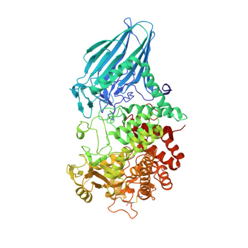
    Proteins belonging to glycoside hydrolase family 63 (GH63) are found in bacteria, archaea and eukaryotes. Although the eukaryotic GH63 proteins have been identified as processing α-glucosidase I, the substrate specificities of the bacterial and archaeal GH63 proteins are not clear. Here, we converted a bacterial GH63 enzyme, Escherichia coli YgjK, to a glycosynthase to probe its substrate specificity. Two mutants of YgjK (E727A and D324N) were constructed, and both mutants showed glycosynthase activity. The reactions of E727A with β-D-glucosyl fluoride and monosaccharides showed that the largest amount of glycosynthase product accumulated when galactose was employed as an acceptor molecule. The crystal structure of E727A complexed with the reaction product indicated that the disaccharide bound at the active site was 2-O-α-D-glucopyranosyl-α-D-galactopyranose (Glc12Gal). A comparison of the structures of E727A-Glc12Gal and D324N-melibiose showed that there were two main types of conformation: the open and closed forms. The structure of YgjK adopted the closed form when subsite -1 was occupied by glucose. These results suggest that sugars containing the Glc12Gal structure are the most likely candidates for natural substrates of YgjK.


  • Organizational Affiliation

    Department of Applied Biological Science, Tokyo University of Agriculture and Technology, Fuchu, Tokyo, Japan.


Macromolecules
Find similar proteins by:  (by identity cutoff)  |  3D Structure
Entity ID: 1
MoleculeChains Sequence LengthOrganismDetailsImage
Uncharacterized protein YgjK
A, B
760Escherichia coli K-12Mutation(s): 1 
Gene Names: b3080JW3051ygjK
UniProt
Find proteins for P42592 (Escherichia coli (strain K12))
Explore P42592 
Go to UniProtKB:  P42592
Entity Groups  
Sequence Clusters30% Identity50% Identity70% Identity90% Identity95% Identity100% Identity
UniProt GroupP42592
Sequence Annotations
Expand
  • Reference Sequence
Oligosaccharides

Help

Entity ID: 2
MoleculeChains Length2D Diagram Glycosylation3D Interactions
alpha-D-glucopyranose-(1-2)-alpha-D-galactopyranose
C, D
2N/A
Glycosylation Resources
GlyTouCan:  G18041KS
GlyCosmos:  G18041KS
GlyGen:  G18041KS
Experimental Data & Validation

Experimental Data

  • Method: X-RAY DIFFRACTION
  • Resolution: 2.00 Å
  • R-Value Free: 0.226 
  • R-Value Work: 0.174 
  • R-Value Observed: 0.176 
  • Space Group: P 1 21 1
Unit Cell:
Length ( Å )Angle ( ˚ )
a = 58.884α = 90
b = 138.136β = 99.03
c = 82.547γ = 90
Software Package:
Software NamePurpose
ADSCdata collection
MOLREPphasing
REFMACrefinement
HKL-2000data reduction
HKL-2000data scaling

Structure Validation

View Full Validation Report


Entry History 

Deposition Data

Revision History  (Full details and data files)

  • Version 1.0: 2013-07-17
    Type: Initial release
  • Version 1.1: 2014-03-12
    Changes: Database references
  • Version 2.0: 2020-07-29
    Type: Remediation
    Reason: Carbohydrate remediation
    Changes: Atomic model, Data collection, Database references, Derived calculations, Structure summary
  • Version 2.1: 2023-11-08
    Changes: Data collection, Database references, Refinement description, Structure summary