3W6V

Crystal structure of the DNA-binding domain of AdpA, the global transcriptional factor, in complex with a target DNA


Experimental Data Snapshot

  • Method: X-RAY DIFFRACTION
  • Resolution: 2.95 Å
  • R-Value Free: 0.278 
  • R-Value Work: 0.236 
  • R-Value Observed: 0.238 

wwPDB Validation   3D Report Full Report


This is version 1.2 of the entry. See complete history


Literature

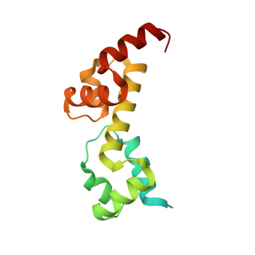
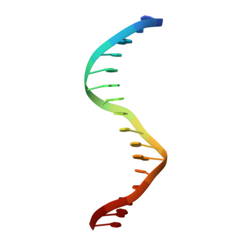
Complex Structure of the DNA-binding Domain of AdpA, the Global Transcription Factor in Streptomyces griseus, and a Target Duplex DNA Reveals the Structural Basis of Its Tolerant DNA Sequence Specificity

Yao, M.D.Ohtsuka, J.Nagata, K.Miyazono, K.Zhi, Y.Ohnishi, Y.Tanokura, M.

(2013) J Biol Chem 288: 31019-31029

  • DOI: https://doi.org/10.1074/jbc.M113.473611
  • Primary Citation of Related Structures:  
    3W6V

  • PubMed Abstract: 

    AdpA serves as the global transcription factor in the A-factor regulatory cascade, controlling the secondary metabolism and morphological differentiation of the filamentous bacterium Streptomyces griseus. AdpA binds to over 500 operator regions with the consensus sequence 5'-TGGCSNGWWY-3' (where S is G or C, W is A or T, Y is T or C, and N is any nucleotide). However, it is still obscure how AdpA can control hundreds of genes. To elucidate the structural basis of this tolerant DNA recognition by AdpA, we focused on the interaction between the DNA-binding domain of AdpA (AdpA-DBD), which consists of two helix-turn-helix motifs, and a target duplex DNA containing the consensus sequence 5'-TGGCGGGTTC-3'. The crystal structure of the AdpA-DBD-DNA complex and the mutant analysis of AdpA-DBD revealed its unique manner of DNA recognition, whereby only two arginine residues directly recognize the consensus sequence, explaining the strict recognition of G and C at positions 2 and 4, respectively, and the tolerant recognition of other positions of the consensus sequence. AdpA-DBD confers tolerant DNA sequence specificity to AdpA, allowing it to control hundreds of genes as a global transcription factor.


  • Organizational Affiliation

    From the Departments of Applied Biological Chemistry and.


Macromolecules

Find similar proteins by:  (by identity cutoff)  |  3D Structure
Entity ID: 1
MoleculeChains Sequence LengthOrganismDetailsImage
AdpA147Streptomyces griseusMutation(s): 0 
Gene Names: adpA
UniProt
Find proteins for Q9S166 (Streptomyces griseus)
Explore Q9S166 
Go to UniProtKB:  Q9S166
Entity Groups  
Sequence Clusters30% Identity50% Identity70% Identity90% Identity95% Identity100% Identity
UniProt GroupQ9S166
Sequence Annotations
Expand
  • Reference Sequence

Find similar nucleic acids by:  Sequence   |   3D Structure  

Entity ID: 2
MoleculeChains LengthOrganismImage
DNA (5'-D(*CP*TP*GP*TP*GP*AP*AP*CP*CP*CP*GP*CP*CP*AP*AP*C)-3')16N/A
Sequence Annotations
Expand
  • Reference Sequence

Find similar nucleic acids by:  Sequence   |   3D Structure  

Entity ID: 3
MoleculeChains LengthOrganismImage
DNA (5'-D(*AP*GP*GP*TP*TP*GP*GP*CP*GP*GP*GP*TP*TP*CP*AP*C)-3')16N/A
Sequence Annotations
Expand
  • Reference Sequence
Experimental Data & Validation

Experimental Data

  • Method: X-RAY DIFFRACTION
  • Resolution: 2.95 Å
  • R-Value Free: 0.278 
  • R-Value Work: 0.236 
  • R-Value Observed: 0.238 
  • Space Group: C 2 2 21
Unit Cell:
Length ( Å )Angle ( ˚ )
a = 76.51α = 90
b = 100.64β = 90
c = 100.91γ = 90
Software Package:
Software NamePurpose
ADSCdata collection
XDSdata scaling
REFMACrefinement
HKL-2000data reduction

Structure Validation

View Full Validation Report


Entry History 

Deposition Data

Revision History  (Full details and data files)

  • Version 1.0: 2013-09-18
    Type: Initial release
  • Version 1.1: 2013-11-20
    Changes: Database references
  • Version 1.2: 2023-11-08
    Changes: Data collection, Database references, Refinement description