


Crystal structure of a glycoside hydrolase
Oyama, T., Schmitz, G.E., Dodd, D., Han, Y., Burnett, A., Nagasawa, N., Mackie, R.I., Nakamura, H., Morikawa, K., Cann, I.K.O.To be published.
Experimental Data Snapshot
wwPDB Validation 3D Report Full Report

Entity ID: 1 | |||||
|---|---|---|---|---|---|
| Molecule | Chains | Sequence Length | Organism | Details | Image |
| Bifunctional endomannanase/endoglucanase | 330 | Caldanaerobius polysaccharolyticus | Mutation(s): 0 Gene Names: man5B | 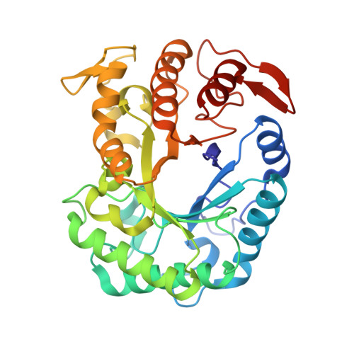 | |
UniProt | |||||
Find proteins for D9J0D7 (Caldanaerobius polysaccharolyticus) Explore D9J0D7 Go to UniProtKB: D9J0D7 | |||||
Entity Groups | |||||
| Sequence Clusters | 30% Identity50% Identity70% Identity90% Identity95% Identity100% Identity | ||||
| UniProt Group | D9J0D7 | ||||
Sequence AnnotationsExpand | |||||
| |||||
| Ligands 1 Unique | |||||
|---|---|---|---|---|---|
| ID | Chains | Name / Formula / InChI Key | 2D Diagram | 3D Interactions | |
| TRS Query on TRS | C [auth A], D [auth B] | 2-AMINO-2-HYDROXYMETHYL-PROPANE-1,3-DIOL C4 H12 N O3 LENZDBCJOHFCAS-UHFFFAOYSA-O |  | ||
| Length ( Å ) | Angle ( ˚ ) |
|---|---|
| a = 50.133 | α = 90 |
| b = 147.642 | β = 104.51 |
| c = 55.422 | γ = 90 |
| Software Name | Purpose |
|---|---|
| ADSC | data collection |
| CNS | refinement |
| HKL-2000 | data reduction |
| HKL-2000 | data scaling |
| CNS | phasing |
RCSB PDB (citation) is hosted by
RCSB PDB is a member of the

