3VYL

Structure of L-ribulose 3-epimerase


Experimental Data Snapshot

  • Method: X-RAY DIFFRACTION
  • Resolution: 2.70 Å
  • R-Value Free: 0.264 
  • R-Value Work: 0.209 
  • R-Value Observed: 0.212 

wwPDB Validation   3D Report Full Report


This is version 1.2 of the entry. See complete history


Literature

Structural insight into L-ribulose 3-epimerase from Mesorhizobium loti.

Uechi, K.Sakuraba, H.Yoshihara, A.Morimoto, K.Takata, G.

(2013) Acta Crystallogr D Biol Crystallogr 69: 2330-2339

  • DOI: https://doi.org/10.1107/S0907444913021665
  • Primary Citation of Related Structures:  
    3VYL

  • PubMed Abstract: 

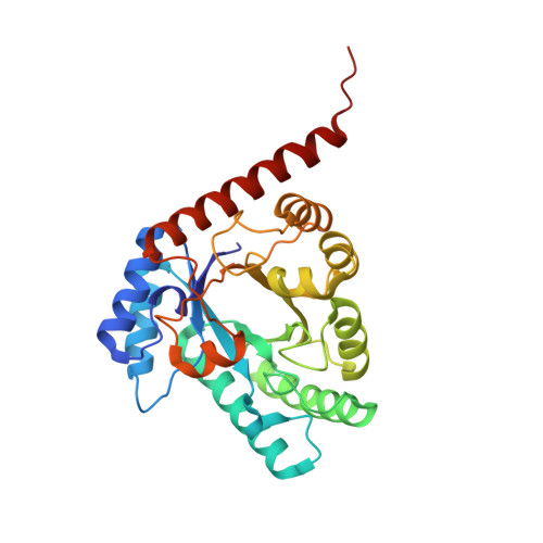
    L-Ribulose 3-epimerase (L-RE) from Mesorhizobium loti has been identified as the first ketose 3-epimerase that shows the highest observed activity towards ketopentoses. In the present study, the crystal structure of the enzyme was determined to 2.7 Å resolution. The asymmetric unit contained two homotetramers with the monomer folded into an (α/β)8-barrel carrying four additional short α-helices. The overall structure of M. loti L-RE showed significant similarity to the structures of ketose 3-epimerases from Pseudomonas cichorii, Agrobacterium tumefaciens and Clostridium cellulolyticum, which use ketohexoses as preferred substrates. However, the size of the C-terminal helix (α8) was much larger in M. loti L-RE than the corresponding helices in the other enzymes. In M. loti L-RE the α8 helix and the following C-terminal tail possessed a unique subunit-subunit interface which promoted the formation of additional intermolecular interactions and strengthened the enzyme stability. Structural comparisons revealed that the relatively small hydrophobic pocket of the enzyme around the substrate was likely to be the main factor responsible for the marked specificity for ketopentoses shown by M. loti L-RE.


  • Organizational Affiliation

    Rare Sugar Research Center, Kagawa University, 2393 Ikenobe, Miki-cho, Kita-gun, Kagawa 761-0795, Japan.


Macromolecules
Find similar proteins by:  (by identity cutoff)  |  3D Structure
Entity ID: 1
MoleculeChains Sequence LengthOrganismDetailsImage
L-ribulose 3-epimerase
A, B, C, D, E
A, B, C, D, E, F, G, H
297Mesorhizobium lotiMutation(s): 0 
EC: 5.3.1
UniProt
Find proteins for N0DP12 (Rhizobium loti)
Explore N0DP12 
Go to UniProtKB:  N0DP12
Entity Groups  
Sequence Clusters30% Identity50% Identity70% Identity90% Identity95% Identity100% Identity
UniProt GroupN0DP12
Sequence Annotations
Expand
  • Reference Sequence
Small Molecules
Ligands 2 Unique
IDChains Name / Formula / InChI Key2D Diagram3D Interactions
MPD
Query on MPD

Download Ideal Coordinates CCD File 
J [auth A],
L [auth B],
O [auth D],
Q [auth E]
(4S)-2-METHYL-2,4-PENTANEDIOL
C6 H14 O2
SVTBMSDMJJWYQN-YFKPBYRVSA-N
MN
Query on MN

Download Ideal Coordinates CCD File 
I [auth A]
K [auth B]
M [auth C]
N [auth D]
P [auth E]
I [auth A],
K [auth B],
M [auth C],
N [auth D],
P [auth E],
R [auth F],
S [auth G],
T [auth H]
MANGANESE (II) ION
Mn
WAEMQWOKJMHJLA-UHFFFAOYSA-N
Experimental Data & Validation

Experimental Data

  • Method: X-RAY DIFFRACTION
  • Resolution: 2.70 Å
  • R-Value Free: 0.264 
  • R-Value Work: 0.209 
  • R-Value Observed: 0.212 
  • Space Group: C 2 2 21
Unit Cell:
Length ( Å )Angle ( ˚ )
a = 202.511α = 90
b = 206.234β = 90
c = 121.975γ = 90
Software Package:
Software NamePurpose
HKL-2000data collection
MOLREPphasing
REFMACrefinement
HKL-2000data reduction
HKL-2000data scaling

Structure Validation

View Full Validation Report


Entry History 

Deposition Data

Revision History  (Full details and data files)

  • Version 1.0: 2013-10-02
    Type: Initial release
  • Version 1.1: 2013-12-18
    Changes: Database references
  • Version 1.2: 2023-11-08
    Changes: Data collection, Database references, Derived calculations, Refinement description