


Crystal Structure of Xylooligosaccharide-Binding Protein from Streptomyces thermoviolaceus OPC-520: Dramatic Conformational Change with Ligand binding
Tomoo, K., Ishida, T., Miyamoto, K., Tsujibo, H.To be published.
Experimental Data Snapshot
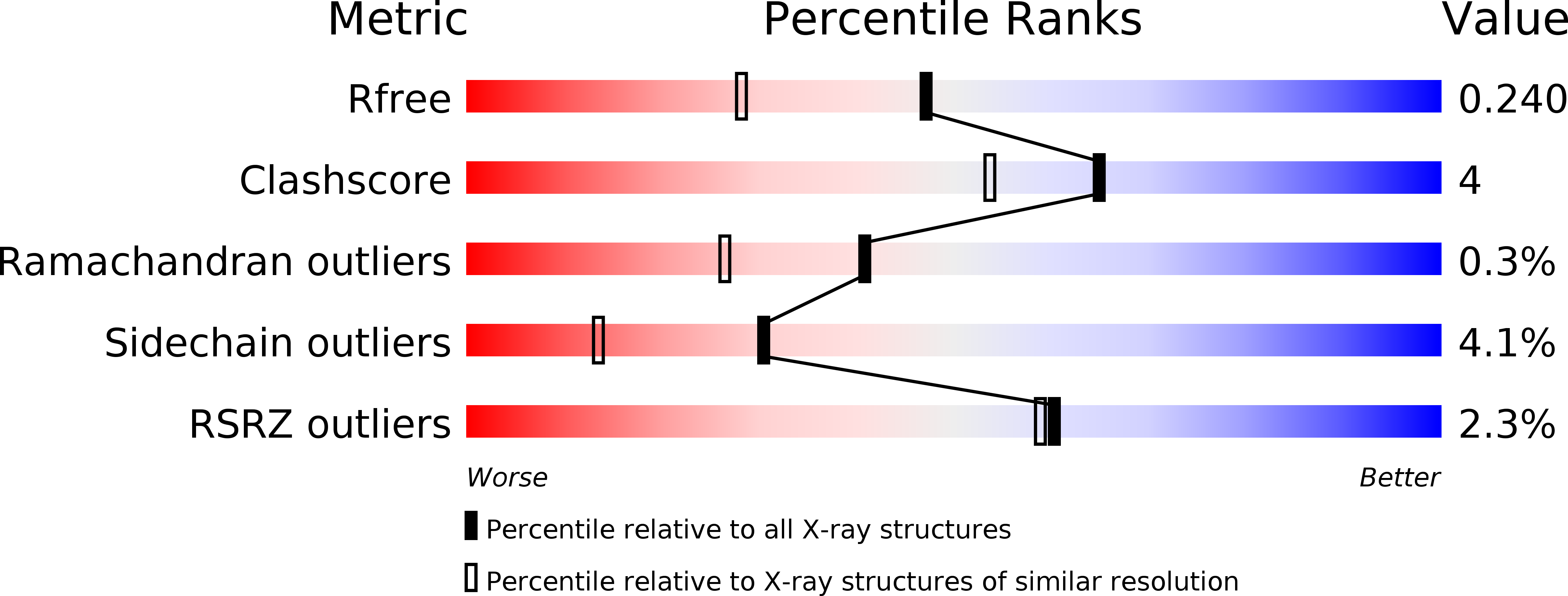
wwPDB Validation 3D Report Full Report
Entity ID: 1 | |||||
|---|---|---|---|---|---|
| Molecule | Chains | Sequence Length | Organism | Details | Image |
| Putative sugar-binding lipoprotein | 436 | Streptomyces thermoviolaceus | Mutation(s): 0 Gene Names: bxlD | 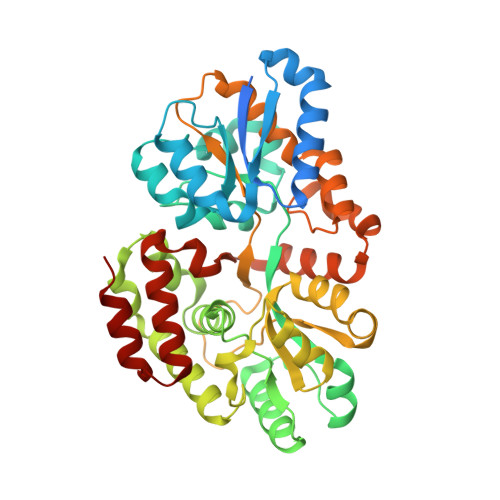 | |
UniProt | |||||
Find proteins for Q76BU9 (Streptomyces thermoviolaceus) Explore Q76BU9 Go to UniProtKB: Q76BU9 | |||||
Entity Groups | |||||
| Sequence Clusters | 30% Identity50% Identity70% Identity90% Identity95% Identity100% Identity | ||||
| UniProt Group | Q76BU9 | ||||
Sequence AnnotationsExpand | |||||
| |||||
| Ligands 1 Unique | |||||
|---|---|---|---|---|---|
| ID | Chains | Name / Formula / InChI Key | 2D Diagram | 3D Interactions | |
| GOL Query on GOL | B [auth A], C [auth A], D [auth A], E [auth A] | GLYCEROL C3 H8 O3 PEDCQBHIVMGVHV-UHFFFAOYSA-N |  | ||
| Length ( Å ) | Angle ( ˚ ) |
|---|---|
| a = 44.634 | α = 90 |
| b = 63.272 | β = 103.05 |
| c = 66.402 | γ = 90 |
| Software Name | Purpose |
|---|---|
| CrystalClear | data collection |
| SOLVE | phasing |
| REFMAC | refinement |
| CrystalClear | data reduction |
| CrystalClear | data scaling |
RCSB PDB (citation) is hosted by
RCSB PDB is a member of the

