3VSL

Crystal structure of penicillin-binding protein 3 (PBP3) from methicilin-resistant Staphylococcus aureus in the cefotaxime bound form.


Experimental Data Snapshot

  • Method: X-RAY DIFFRACTION
  • Resolution: 2.40 Å
  • R-Value Free: 0.316 
  • R-Value Work: 0.247 
  • R-Value Observed: 0.250 

wwPDB Validation   3D Report Full Report


Ligand Structure Quality Assessment 


This is version 1.1 of the entry. See complete history


Literature

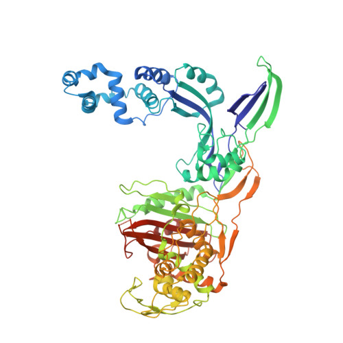
Crystal Structures of Penicillin-Binding Protein 3 (PBP3) from Methicillin-Resistant Staphylococcus aureus in the Apo and Cefotaxime-Bound Forms.

Yoshida, H.Kawai, F.Obayashi, E.Akashi, S.Roper, D.I.Tame, J.R.Park, S.Y.

(2012) J Mol Biol 423: 351-364

  • DOI: https://doi.org/10.1016/j.jmb.2012.07.012
  • Primary Citation of Related Structures:  
    3VSK, 3VSL

  • PubMed Abstract: 

    Staphylococcus aureus is a widespread Gram-positive opportunistic pathogen, and a methicillin-resistant form (MRSA) is particularly difficult to treat clinically. We have solved two crystal structures of penicillin-binding protein (PBP) 3 (PBP3) from MRSA, the apo form and a complex with the β-lactam antibiotic cefotaxime, and used electrospray mass spectrometry to measure its sensitivity to a variety of penicillin derivatives. PBP3 is a class B PBP, possessing an N-terminal non-penicillin-binding domain, sometimes called a dimerization domain, and a C-terminal transpeptidase domain. The model shows a different orientation of its two domains compared to earlier models of other class B PBPs and a novel, larger N-domain. Consistent with the nomenclature of "dimerization domain", the N-terminal region forms an apparently tight interaction with a neighboring molecule related by a 2-fold symmetry axis in the crystal structure. This dimer form is predicted to be highly stable in solution by the PISA server, but mass spectrometry and analytical ultracentrifugation provide unequivocal evidence that the protein is a monomer in solution.


  • Organizational Affiliation

    Protein Design Laboratory, Yokohama City University, Suehiro 1-7-29, Tsurumi-ku, Yokohama 230-0045, Japan.


Macromolecules
Find similar proteins by:  (by identity cutoff)  |  3D Structure
Entity ID: 1
MoleculeChains Sequence LengthOrganismDetailsImage
Penicillin-binding protein 3
A, B
646Staphylococcus aureus subsp. aureus MW2Mutation(s): 0 
Gene Names: pbp3MW1504
UniProt
Find proteins for A0A0H3K0B9 (Staphylococcus aureus (strain MW2))
Explore A0A0H3K0B9 
Go to UniProtKB:  A0A0H3K0B9
Entity Groups  
Sequence Clusters30% Identity50% Identity70% Identity90% Identity95% Identity100% Identity
UniProt GroupA0A0H3K0B9
Sequence Annotations
Expand
  • Reference Sequence
Small Molecules
Ligands 1 Unique
IDChains Name / Formula / InChI Key2D Diagram3D Interactions
CEF
Query on CEF

Download Ideal Coordinates CCD File 
C [auth A],
D [auth B]
CEFOTAXIME, C3' cleaved, open, bound form
C14 H15 N5 O5 S2
NRYMPLKBKFIWQC-YVCCLBOHSA-N
Experimental Data & Validation

Experimental Data

  • Method: X-RAY DIFFRACTION
  • Resolution: 2.40 Å
  • R-Value Free: 0.316 
  • R-Value Work: 0.247 
  • R-Value Observed: 0.250 
  • Space Group: P 43 21 2
Unit Cell:
Length ( Å )Angle ( ˚ )
a = 143.031α = 90
b = 143.031β = 90
c = 189.61γ = 90
Software Package:
Software NamePurpose
HKL-2000data collection
MOLREPphasing
PHENIXrefinement
DENZOdata reduction
SCALEPACKdata scaling

Structure Validation

View Full Validation Report


Ligand Structure Quality Assessment 


Entry History 

Deposition Data

Revision History  (Full details and data files)

  • Version 1.0: 2012-10-31
    Type: Initial release
  • Version 1.1: 2017-03-15
    Changes: Non-polymer description