3UOM

Ca2+ complex of Human skeletal calsequestrin


Experimental Data Snapshot

  • Method: X-RAY DIFFRACTION
  • Resolution: 2.02 Å
  • R-Value Free: 0.213 
  • R-Value Work: 0.182 
  • R-Value Observed: 0.182 

wwPDB Validation   3D Report Full Report


This is version 1.3 of the entry. See complete history


Literature

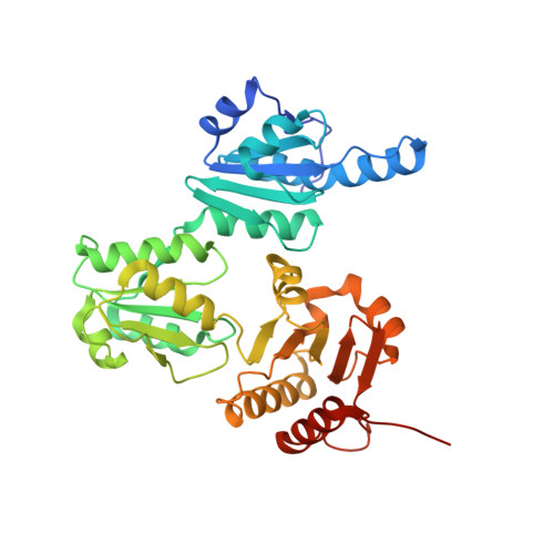
High-capacity Ca2+ Binding of Human Skeletal Calsequestrin.

Sanchez, E.J.Lewis, K.M.Danna, B.R.Kang, C.

(2012) J Biol Chem 287: 11592-11601

  • DOI: https://doi.org/10.1074/jbc.M111.335075
  • Primary Citation of Related Structures:  
    3TRP, 3UOM

  • PubMed Abstract: 

    Calsequestrin, the major calcium storage protein in both cardiac and skeletal muscle, binds large amounts of Ca(2+) in the sarcoplasmic reticulum and releases them during muscle contraction. For the first time, the crystal structures of Ca(2+) complexes for both human (hCASQ1) and rabbit (rCASQ1) skeletal calsequestrin were determined, clearly defining their Ca(2+) sequestration capabilities through resolution of high- and low-affinity Ca(2+)-binding sites. rCASQ1 crystallized in low CaCl(2) buffer reveals three high-affinity Ca(2+) sites with trigonal bipyramidal, octahedral, and pentagonal bipyramidal coordination geometries, along with three low-affinity Ca(2+) sites. hCASQ1 crystallized in high CaCl(2) shows 15 Ca(2+) ions, including the six Ca(2+) ions in rCASQ1. Most of the low-affinity sites, some of which are μ-carboxylate-bridged, are established by the rotation of dimer interfaces, indicating cooperative Ca(2+) binding that is consistent with our atomic absorption spectroscopic data. On the basis of these findings, we propose a mechanism for the observed in vitro and in vivo dynamic high-capacity and low-affinity Ca(2+)-binding activity of calsequestrin.


  • Organizational Affiliation

    School of Molecular Biosciences, Washington State University, Pullman, Washington 99164, USA.


Macromolecules
Find similar proteins by:  (by identity cutoff)  |  3D Structure
Entity ID: 1
MoleculeChains Sequence LengthOrganismDetailsImage
Calsequestrin-1
A, B, C, D, E
A, B, C, D, E, F
362Homo sapiensMutation(s): 0 
UniProt & NIH Common Fund Data Resources
Find proteins for P31415 (Homo sapiens)
Explore P31415 
Go to UniProtKB:  P31415
PHAROS:  P31415
GTEx:  ENSG00000143318 
Entity Groups  
Sequence Clusters30% Identity50% Identity70% Identity90% Identity95% Identity100% Identity
UniProt GroupP31415
Sequence Annotations
Expand
  • Reference Sequence
Small Molecules
Ligands 3 Unique
IDChains Name / Formula / InChI Key2D Diagram3D Interactions
MPD
Query on MPD

Download Ideal Coordinates CCD File 
AB [auth C]
BA [auth B]
BB [auth C]
DA [auth B]
FA [auth B]
AB [auth C],
BA [auth B],
BB [auth C],
DA [auth B],
FA [auth B],
GA [auth B],
H [auth A],
I [auth A],
J [auth A],
K [auth A],
KD [auth F],
L [auth A],
LD [auth F],
MD [auth F],
TC [auth E],
ZA [auth C]
(4S)-2-METHYL-2,4-PENTANEDIOL
C6 H14 O2
SVTBMSDMJJWYQN-YFKPBYRVSA-N
MRD
Query on MRD

Download Ideal Coordinates CCD File 
CA [auth B]
CB [auth C]
DB [auth C]
EA [auth B]
G [auth A]
CA [auth B],
CB [auth C],
DB [auth C],
EA [auth B],
G [auth A],
HA [auth B],
ID [auth F],
JD [auth F],
M [auth A],
ND [auth F],
OD [auth F],
PC [auth E],
PD [auth F],
QC [auth E],
RC [auth E],
SC [auth E],
TB [auth D],
UB [auth D],
UC [auth E],
VB [auth D],
WB [auth D],
XA [auth C],
XB [auth D],
YA [auth C],
YB [auth D],
ZB [auth D]
(4R)-2-METHYLPENTANE-2,4-DIOL
C6 H14 O2
SVTBMSDMJJWYQN-RXMQYKEDSA-N
CA
Query on CA

Download Ideal Coordinates CCD File 
AA [auth A]
AC [auth D]
AD [auth E]
AE [auth F]
BC [auth D]
AA [auth A],
AC [auth D],
AD [auth E],
AE [auth F],
BC [auth D],
BD [auth E],
BE [auth F],
CC [auth D],
CD [auth E],
CE [auth F],
DC [auth D],
DD [auth E],
DE [auth F],
EB [auth C],
EC [auth D],
ED [auth E],
FB [auth C],
FC [auth D],
FD [auth E],
GB [auth C],
GC [auth D],
GD [auth E],
HB [auth C],
HC [auth D],
HD [auth E],
IA [auth B],
IB [auth C],
IC [auth D],
JA [auth B],
JB [auth C],
JC [auth D],
KA [auth B],
KB [auth C],
KC [auth D],
LA [auth B],
LB [auth C],
LC [auth D],
MA [auth B],
MB [auth C],
MC [auth D],
N [auth A],
NA [auth B],
NB [auth C],
NC [auth D],
O [auth A],
OA [auth B],
OB [auth C],
OC [auth D],
P [auth A],
PA [auth B],
PB [auth C],
Q [auth A],
QA [auth B],
QB [auth C],
QD [auth F],
R [auth A],
RA [auth B],
RB [auth C],
RD [auth F],
S [auth A],
SA [auth B],
SB [auth C],
SD [auth F],
T [auth A],
TA [auth B],
TD [auth F],
U [auth A],
UA [auth B],
UD [auth F],
V [auth A],
VA [auth B],
VC [auth E],
VD [auth F],
W [auth A],
WA [auth B],
WC [auth E],
WD [auth F],
X [auth A],
XC [auth E],
XD [auth F],
Y [auth A],
YC [auth E],
YD [auth F],
Z [auth A],
ZC [auth E],
ZD [auth F]
CALCIUM ION
Ca
BHPQYMZQTOCNFJ-UHFFFAOYSA-N
Experimental Data & Validation

Experimental Data

  • Method: X-RAY DIFFRACTION
  • Resolution: 2.02 Å
  • R-Value Free: 0.213 
  • R-Value Work: 0.182 
  • R-Value Observed: 0.182 
  • Space Group: P 1
Unit Cell:
Length ( Å )Angle ( ˚ )
a = 89.799α = 90.13
b = 89.793β = 89.9
c = 119.161γ = 60.05
Software Package:
Software NamePurpose
PHENIXrefinement

Structure Validation

View Full Validation Report


Entry History 

Deposition Data

Revision History  (Full details and data files)

  • Version 1.0: 2012-02-22
    Type: Initial release
  • Version 1.1: 2012-04-25
    Changes: Database references
  • Version 1.2: 2012-07-18
    Changes: Database references
  • Version 1.3: 2023-09-13
    Changes: Data collection, Database references, Derived calculations, Refinement description