3UD0

ATP synthase C10 ring in proton-unlocked conformation at PH 5.5


Experimental Data Snapshot

  • Method: X-RAY DIFFRACTION
  • Resolution: 2.00 Å
  • R-Value Free: 0.221 
  • R-Value Work: 0.200 
  • R-Value Observed: 0.201 

wwPDB Validation   3D Report Full Report


This is version 1.3 of the entry. See complete history


Literature

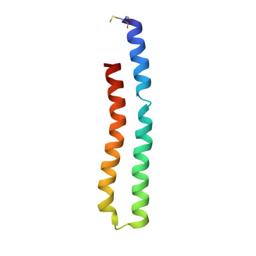
Structure of the c(10) ring of the yeast mitochondrial ATP synthase in the open conformation.

Symersky, J.Pagadala, V.Osowski, D.Krah, A.Meier, T.Faraldo-Gomez, J.D.Mueller, D.M.

(2012) Nat Struct Mol Biol 19: 485-491

  • DOI: https://doi.org/10.1038/nsmb.2284
  • Primary Citation of Related Structures:  
    3U2F, 3U2Y, 3U32, 3UD0

  • PubMed Abstract: 

    The proton pore of the F(1)F(o) ATP synthase consists of a ring of c subunits, which rotates, driven by downhill proton diffusion across the membrane. An essential carboxylate side chain in each subunit provides a proton-binding site. In all the structures of c-rings reported to date, these sites are in a closed, ion-locked state. Structures are here presented of the c(10) ring from Saccharomyces cerevisiae determined at pH 8.3, 6.1 and 5.5, at resolutions of 2.0 Å, 2.5 Å and 2.0 Å, respectively. The overall structure of this mitochondrial c-ring is similar to known homologs, except that the essential carboxylate, Glu59, adopts an open extended conformation. Molecular dynamics simulations reveal that opening of the essential carboxylate is a consequence of the amphiphilic nature of the crystallization buffer. We propose that this new structure represents the functionally open form of the c subunit, which facilitates proton loading and release.


  • Organizational Affiliation

    Department of Biochemistry and Molecular Biology, Rosalind Franklin University of Medicine and Science, The Chicago Medical School, Chicago, Illinois, USA.


Macromolecules
Find similar proteins by:  (by identity cutoff)  |  3D Structure
Entity ID: 1
MoleculeChains Sequence LengthOrganismDetailsImage
ATP synthase subunit C, mitochondrialA [auth K],
B [auth L],
C [auth M],
D [auth N],
E [auth O]
76Saccharomyces cerevisiaeMutation(s): 0 
Membrane Entity: Yes 
UniProt
Find proteins for P61829 (Saccharomyces cerevisiae (strain ATCC 204508 / S288c))
Explore P61829 
Go to UniProtKB:  P61829
Entity Groups  
Sequence Clusters30% Identity50% Identity70% Identity90% Identity95% Identity100% Identity
UniProt GroupP61829
Sequence Annotations
Expand
  • Reference Sequence
Small Molecules
Modified Residues  1 Unique
IDChains TypeFormula2D DiagramParent
FME
Query on FME
A [auth K],
B [auth L],
C [auth M],
D [auth N],
E [auth O]
L-PEPTIDE LINKINGC6 H11 N O3 SMET
Experimental Data & Validation

Experimental Data

  • Method: X-RAY DIFFRACTION
  • Resolution: 2.00 Å
  • R-Value Free: 0.221 
  • R-Value Work: 0.200 
  • R-Value Observed: 0.201 
  • Space Group: P 42 2 2
Unit Cell:
Length ( Å )Angle ( ˚ )
a = 54.159α = 90
b = 54.159β = 90
c = 245.392γ = 90
Software Package:
Software NamePurpose
Blu-Icedata collection
REFMACrefinement
HKL-2000data reduction
HKL-2000data scaling
REFMACphasing

Structure Validation

View Full Validation Report


Entry History 

Deposition Data

Revision History  (Full details and data files)

  • Version 1.0: 2012-02-08
    Type: Initial release
  • Version 1.1: 2012-04-18
    Changes: Database references
  • Version 1.2: 2012-06-13
    Changes: Database references
  • Version 1.3: 2023-09-13
    Changes: Data collection, Database references, Derived calculations, Refinement description