3UAQ

Crystal Structure of the N-lobe Domain of Lactoferrin Binding Protein B (LbpB) of Moraxella bovis


Experimental Data Snapshot

  • Method: X-RAY DIFFRACTION
  • Resolution: 2.93 Å
  • R-Value Free: 0.269 
  • R-Value Work: 0.220 
  • R-Value Observed: 0.223 

wwPDB Validation   3D Report Full Report


This is version 1.4 of the entry. See complete history


Literature

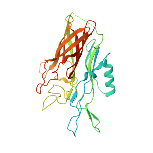
Crystal structure of the N-lobe of lactoferrin binding protein B from Moraxella bovis(1).

Arutyunova, E.Brooks, C.L.Beddek, A.Mak, M.W.Schryvers, A.B.Lemieux, M.J.

(2012) Biochem Cell Biol 90: 351-361

  • DOI: https://doi.org/10.1139/o11-078
  • Primary Citation of Related Structures:  
    3UAQ

  • PubMed Abstract: 

    Lactoferrin (Lf) is a bi-lobed, iron-binding protein found on mucosal surfaces and at sites of inflammation. Gram-negative pathogens from the Neisseriaceae and Moraxellaceae families are capable of using Lf as a source of iron for growth through a process mediated by a bacterial surface receptor that directly binds host Lf. This receptor consists of an integral outer membrane protein, lactoferrin binding protein A (LbpA), and a surface lipoprotein, lactoferrin binding protein B (LbpB). The N-lobe of the homologous transferrin binding protein B, TbpB, has been shown to facilitate transferrin binding in the process of iron acquisition. Currently there is little known about the role of LbpB in iron acquisition or how Lf interacts with the bacterial receptor proteins. No structural information on any LbpB or domain is available. In this study, we express and purify from Escherichia coli the full-length LbpB and the N-lobe of LbpB from the bovine pathogen Moraxella bovis for crystallization trials. We demonstrate that M. bovis LbpB binds to bovine but not human Lf. We also report the crystal structure of the N-terminal lobe of LbpB from M. bovis and compare it with the published structures of TbpB to speculate on the process of Lf mediated iron acquisition.


  • Organizational Affiliation

    Department of Biochemistry, University of Alberta, Edmonton, AB T6G 2H7, Canada.


Macromolecules
Find similar proteins by:  (by identity cutoff)  |  3D Structure
Entity ID: 1
MoleculeChains Sequence LengthOrganismDetailsImage
LbpB B-lobe
A, B
341Moraxella bovisMutation(s): 0 
Gene Names: LbpB
UniProt
Find proteins for H9KVH9 (Moraxella bovis)
Explore H9KVH9 
Go to UniProtKB:  H9KVH9
Entity Groups  
Sequence Clusters30% Identity50% Identity70% Identity90% Identity95% Identity100% Identity
UniProt GroupH9KVH9
Sequence Annotations
Expand
  • Reference Sequence
Experimental Data & Validation

Experimental Data

  • Method: X-RAY DIFFRACTION
  • Resolution: 2.93 Å
  • R-Value Free: 0.269 
  • R-Value Work: 0.220 
  • R-Value Observed: 0.223 
  • Space Group: P 1 21 1
Unit Cell:
Length ( Å )Angle ( ˚ )
a = 42.599α = 90
b = 83.928β = 101.11
c = 98.589γ = 90
Software Package:
Software NamePurpose
DENZOdata reduction
SCALEPACKdata scaling
PHENIXrefinement
PDB_EXTRACTdata extraction
HKL-2000data reduction

Structure Validation

View Full Validation Report


Entry History 

Deposition Data

Revision History  (Full details and data files)

  • Version 1.0: 2012-03-28
    Type: Initial release
  • Version 1.1: 2012-05-30
    Changes: Database references
  • Version 1.2: 2017-11-08
    Changes: Refinement description, Structure summary
  • Version 1.3: 2018-01-24
    Changes: Structure summary
  • Version 1.4: 2023-09-13
    Changes: Data collection, Database references, Refinement description