Structural insights into the catalytic mechanism of Escherichia coli selenophosphate synthetase.
Noinaj, N., Wattanasak, R., Lee, D.Y., Wally, J.L., Piszczek, G., Chock, P.B., Stadtman, T.C., Buchanan, S.K.(2012) J Bacteriol 194: 499-508
- PubMed: 22081394
- DOI: https://doi.org/10.1128/JB.06012-11
- Primary Citation of Related Structures:
3U0O - PubMed Abstract:
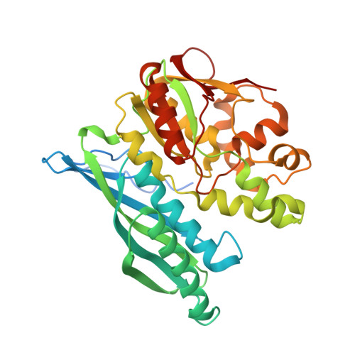
Selenophosphate synthetase (SPS) catalyzes the synthesis of selenophosphate, the selenium donor for the biosynthesis of selenocysteine and 2-selenouridine residues in seleno-tRNA. Selenocysteine, known as the 21st amino acid, is then incorporated into proteins during translation to form selenoproteins which serve a variety of cellular processes. SPS activity is dependent on both Mg(2+) and K(+) and uses ATP, selenide, and water to catalyze the formation of AMP, orthophosphate, and selenophosphate. In this reaction, the gamma phosphate of ATP is transferred to the selenide to form selenophosphate, while ADP is hydrolyzed to form orthophosphate and AMP. Most of what is known about the function of SPS has derived from studies investigating Escherichia coli SPS (EcSPS) as a model system. Here we report the crystal structure of the C17S mutant of SPS from E. coli (EcSPS(C17S)) in apo form (without ATP bound). EcSPS(C17S) crystallizes as a homodimer, which was further characterized by analytical ultracentrifugation experiments. The glycine-rich N-terminal region (residues 1 through 47) was found in the open conformation and was mostly ordered in both structures, with a magnesium cofactor bound at the active site of each monomer involving conserved aspartate residues. Mutating these conserved residues (D51, D68, D91, and D227) along with N87, also found at the active site, to alanine completely abolished AMP production in our activity assays, highlighting their essential role for catalysis in EcSPS. Based on the structural and biochemical analysis of EcSPS reported here and using information obtained from similar studies done with SPS orthologs from Aquifex aeolicus and humans, we propose a catalytic mechanism for EcSPS-mediated selenophosphate synthesis.
Organizational Affiliation:
National Institute of Diabetes and Digestive and Kidney Diseases, National Institutes of Health, Bethesda, Maryland, USA. noinajn@niddk.nih.gov