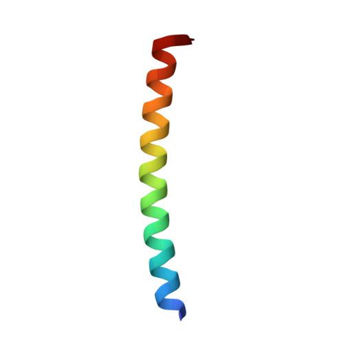
3TQ2

Merohedral twinning in protein crystals revealed a new synthetic three helix bundle motif


Experimental Data Snapshot

  • Method: X-RAY DIFFRACTION
  • Resolution: 1.10 Å
  • R-Value Free: 0.185 
  • R-Value Work: 0.158 
  • R-Value Observed: 0.188 

wwPDB Validation   3D Report Full Report


This is version 1.2 of the entry. See complete history


Literature

Analysis of the crystal structure of a parallel three-stranded coiled coil.

de March, M.Hickey, N.Geremia, S.

(2023) Proteins 91: 1254-1260

  • DOI: https://doi.org/10.1002/prot.26557
  • Primary Citation of Related Structures:  
    3TQ2

  • PubMed Abstract: 

    Here, we present the crystal structure of the synthetic peptide KE1, which contains four K-coil heptads separated in the middle by the QFLMLMF heptad. The structure determination reveals the presence of a canonical parallel three stranded coiled coil. The geometric characteristics of this structure are compared with other coiled coils with the same topology. Furthermore, for this topology, the analysis of the propensity of the single amino acid to occupy a specific position in the heptad sequence is reported. A number of viral proteins use specialized coiled coil tail needles to inject their genetic material into the host cells. The simplicity and regularity of the coiled coil arrangement made it an attractive system for de novo design of key molecules in drug delivery systems, vaccines, and therapeutics.


  • Organizational Affiliation

    Department of Chemical and Pharmaceutical Sciences, University of Trieste, Trieste, Italy.


Macromolecules
Find similar proteins by:  (by identity cutoff)  |  3D Structure
Entity ID: 1
MoleculeChains Sequence LengthOrganismDetailsImage
KE136synthetic constructMutation(s): 0 
Entity Groups  
Sequence Clusters30% Identity50% Identity70% Identity90% Identity95% Identity100% Identity
Sequence Annotations
Expand
  • Reference Sequence
Small Molecules
Ligands 1 Unique
IDChains Name / Formula / InChI Key2D Diagram3D Interactions
SO4
Query on SO4

Download Ideal Coordinates CCD File 
B [auth A]SULFATE ION
O4 S
QAOWNCQODCNURD-UHFFFAOYSA-L
Experimental Data & Validation

Experimental Data

  • Method: X-RAY DIFFRACTION
  • Resolution: 1.10 Å
  • R-Value Free: 0.185 
  • R-Value Work: 0.158 
  • R-Value Observed: 0.188 
  • Space Group: P 3
Unit Cell:
Length ( Å )Angle ( ˚ )
a = 22.365α = 90
b = 22.365β = 90
c = 51.007γ = 120
Software Package:
Software NamePurpose
HKL-2000data collection
AMoREphasing
REFMACrefinement
HKL-2000data reduction
SCALAdata scaling

Structure Validation

View Full Validation Report


Entry History 

Deposition Data

Revision History  (Full details and data files)

  • Version 1.0: 2012-09-26
    Type: Initial release
  • Version 1.1: 2024-01-31
    Changes: Data collection, Database references, Derived calculations
  • Version 1.2: 2024-04-03
    Changes: Refinement description