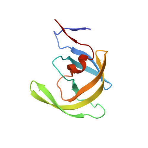
3TOH

HIV-1 Protease - Epoxydic Inhibitor Complex (pH 9 - Orthorombic Crystal form P212121)


Experimental Data Snapshot

  • Method: X-RAY DIFFRACTION
  • Resolution: 1.12 Å
  • R-Value Free: 0.211 
  • R-Value Work: 0.177 
  • R-Value Observed: 0.178 

wwPDB Validation   3D Report Full Report


Ligand Structure Quality Assessment 


This is version 1.5 of the entry. See complete history


Literature

Developing HIV-1 Protease Inhibitors through Stereospecific Reactions in Protein Crystals.

Olajuyigbe, F.M.Demitri, N.De Zorzi, R.Geremia, S.

(2016) Molecules 21

  • DOI: https://doi.org/10.3390/molecules21111458
  • Primary Citation of Related Structures:  
    3TOF, 3TOG, 3TOH

  • PubMed Abstract: 

    Protease inhibitors are key components in the chemotherapy of HIV infection. However, the appearance of viral mutants routinely compromises their clinical efficacy, creating a constant need for new and more potent inhibitors. Recently, a new class of epoxide-based inhibitors of HIV-1 protease was investigated and the configuration of the epoxide carbons was demonstrated to play a crucial role in determining the binding affinity. Here we report the comparison between three crystal structures at near-atomic resolution of HIV-1 protease in complex with the epoxide-based inhibitor, revealing an in-situ epoxide ring opening triggered by a pH change in the mother solution of the crystal. Increased pH in the crystal allows a stereospecific nucleophile attack of an ammonia molecule onto an epoxide carbon, with formation of a new inhibitor containing amino-alcohol functions. The described experiments open a pathway for the development of new stereospecific protease inhibitors from a reactive lead compound.


  • Organizational Affiliation

    Centre of Excellence in Biocrystallography, Department of Chemical and Pharmaceutical Sciences, University of Trieste, Trieste 34127, Italy. folajuyi@futa.edu.ng.


Macromolecules
Find similar proteins by:  (by identity cutoff)  |  3D Structure
Entity ID: 1
MoleculeChains Sequence LengthOrganismDetailsImage
Gag-Pol polyprotein
A, B
99Human immunodeficiency virus type 1 (BRU ISOLATE)Mutation(s): 5 
Gene Names: gag-pol
EC: 3.4.23.16 (PDB Primary Data), 2.7.7.49 (PDB Primary Data), 2.7.7.7 (PDB Primary Data), 3.1.26.13 (PDB Primary Data), 3.1.13.2 (PDB Primary Data)
UniProt
Find proteins for P03367 (Human immunodeficiency virus type 1 group M subtype B (isolate BRU/LAI))
Explore P03367 
Go to UniProtKB:  P03367
Entity Groups  
Sequence Clusters30% Identity50% Identity70% Identity90% Identity95% Identity100% Identity
UniProt GroupP03367
Sequence Annotations
Expand
  • Reference Sequence
Small Molecules
Experimental Data & Validation

Experimental Data

  • Method: X-RAY DIFFRACTION
  • Resolution: 1.12 Å
  • R-Value Free: 0.211 
  • R-Value Work: 0.177 
  • R-Value Observed: 0.178 
  • Space Group: P 21 21 21
Unit Cell:
Length ( Å )Angle ( ˚ )
a = 51.268α = 90
b = 58.377β = 90
c = 61.3γ = 90
Software Package:
Software NamePurpose
HKL-2000data collection
REFMACrefinement
HKL-2000data reduction
HKL-2000data scaling

Structure Validation

View Full Validation Report


Ligand Structure Quality Assessment 


Entry History 

Deposition Data

Revision History  (Full details and data files)

  • Version 1.0: 2012-08-15
    Type: Initial release
  • Version 1.1: 2012-12-12
    Changes: Other
  • Version 1.2: 2014-10-29
    Changes: Structure summary
  • Version 1.3: 2017-11-08
    Changes: Refinement description
  • Version 1.4: 2019-03-20
    Changes: Data collection, Database references
  • Version 1.5: 2023-09-13
    Changes: Data collection, Database references, Derived calculations, Refinement description