3TKG

crystal structure of HIV model protease precursor/saquinavir complex


Experimental Data Snapshot

  • Method: X-RAY DIFFRACTION
  • Resolution: 1.36 Å
  • R-Value Free: 0.214 
  • R-Value Work: 0.160 
  • R-Value Observed: 0.163 

wwPDB Validation   3D Report Full Report


Ligand Structure Quality Assessment 


This is version 1.3 of the entry. See complete history


Literature

Terminal interface conformations modulate dimer stability prior to amino terminal autoprocessing of HIV-1 protease.

Agniswamy, J.Sayer, J.M.Weber, I.T.Louis, J.M.

(2012) Biochemistry 51: 1041-1050

  • DOI: https://doi.org/10.1021/bi201809s
  • Primary Citation of Related Structures:  
    3TKG, 3TKW, 3TL9

  • PubMed Abstract: 

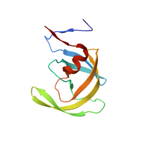
    The HIV-1 protease (PR) mediates its own release (autoprocessing) from the polyprotein precursor, Gag-Pol, flanked by the transframe region (TFR) and reverse transcriptase at its N- and C-termini, respectively. Autoprocessing at the N-terminus of PR mediates stable dimer formation essential for catalytic activity, leading to the formation of infectious virus. An antiparallel β-sheet interface formed by the four N- and C-terminal residues of each subunit is important for dimer stability. Here, we present the first high-resolution crystal structures of model protease precursor-clinical inhibitor (PI darunavir or saquinavir) complexes, revealing varying conformations of the N-terminal flanking (S(-4)FNF(-1)) and interface residues (P(1)QIT(4)). A 180° rotation of the T(4)-L(5) peptide bond is accompanied by a new Q(2)-L(5) hydrogen bond and complete disengagement of PQIT from the β-sheet dimer interface, which may be a feature for intramolecular autoprocessing. This result is consistent with drastically lower thermal stability by 14-20 °C of PI complexes of precursors and the mature PR lacking its PQIT residues (by 18.3 °C). Similar to the TFR-PR precursor, this deletion also results in a darunavir dissociation constant (2 × 10(4))-fold higher and a markedly increased dimer dissociation constant relative to the mature PR. The terminal β-sheet perturbations of the dimeric structure likely account for the drastically poorer inhibition of autoprocessing of TFR-PR relative to the mature PR, even though significant differences in active site-PI interactions in these structures were not observed. The novel conformations of the dimer interface may be exploited to target selectively the protease precursor prior to its N-terminal cleavage.


  • Organizational Affiliation

    Department of Biology, Molecular Basis of Disease Program, Georgia State University, Atlanta, Georgia 30303, United States.


Macromolecules
Find similar proteins by:  (by identity cutoff)  |  3D Structure
Entity ID: 1
MoleculeChains Sequence LengthOrganismDetailsImage
Protease
A, B, C, D
103Human immunodeficiency virus type 1 (BRU ISOLATE)Mutation(s): 0 
Gene Names: gag-pol
EC: 3.4.23.16
UniProt
Find proteins for P03366 (Human immunodeficiency virus type 1 group M subtype B (isolate BH10))
Explore P03366 
Go to UniProtKB:  P03366
Entity Groups  
Sequence Clusters30% Identity50% Identity70% Identity90% Identity95% Identity100% Identity
UniProt GroupP03366
Sequence Annotations
Expand
  • Reference Sequence
Small Molecules
Ligands 3 Unique
IDChains Name / Formula / InChI Key2D Diagram3D Interactions
ROC
Query on ROC

Download Ideal Coordinates CCD File 
G [auth B],
I [auth C]
(2S)-N-[(2S,3R)-4-[(2S,3S,4aS,8aS)-3-(tert-butylcarbamoyl)-3,4,4a,5,6,7,8,8a-octahydro-1H-isoquinolin-2-yl]-3-hydroxy-1 -phenyl-butan-2-yl]-2-(quinolin-2-ylcarbonylamino)butanediamide
C38 H50 N6 O5
QWAXKHKRTORLEM-UGJKXSETSA-N
GOL
Query on GOL

Download Ideal Coordinates CCD File 
F [auth A]GLYCEROL
C3 H8 O3
PEDCQBHIVMGVHV-UHFFFAOYSA-N
CL
Query on CL

Download Ideal Coordinates CCD File 
E [auth A],
H [auth B],
J [auth C],
K [auth D]
CHLORIDE ION
Cl
VEXZGXHMUGYJMC-UHFFFAOYSA-M
Binding Affinity Annotations 
IDSourceBinding Affinity
ROC BindingDB:  3TKG Ki: min: 0.04, max: 138 (nM) from 29 assay(s)
Kd: min: 0.31, max: 67 (nM) from 6 assay(s)
IC50: min: 0.2, max: 270 (nM) from 16 assay(s)
-TΔS: min: -8.03e+1, max: -2.80e+1 (kJ/mol) from 47 assay(s)
ΔH: min: -3.18e+1, max: 43.47 (kJ/mol) from 47 assay(s)
ΔG: min: -6.23e+1, max: -3.55e+1 (kJ/mol) from 44 assay(s)
Biologically Interesting Molecules (External Reference) 1 Unique
Experimental Data & Validation

Experimental Data

  • Method: X-RAY DIFFRACTION
  • Resolution: 1.36 Å
  • R-Value Free: 0.214 
  • R-Value Work: 0.160 
  • R-Value Observed: 0.163 
  • Space Group: P 21 21 21
Unit Cell:
Length ( Å )Angle ( ˚ )
a = 80.074α = 90
b = 88.94β = 90
c = 60.168γ = 90
Software Package:
Software NamePurpose
SERGUIdata collection
PHASERphasing
SHELXL-97refinement
HKL-2000data reduction
HKL-2000data scaling

Structure Validation

View Full Validation Report


Ligand Structure Quality Assessment 


Entry History 

Deposition Data

Revision History  (Full details and data files)

  • Version 1.0: 2012-04-25
    Type: Initial release
  • Version 1.1: 2012-12-12
    Changes: Other
  • Version 1.2: 2017-11-08
    Changes: Refinement description
  • Version 1.3: 2023-09-13
    Changes: Data collection, Database references, Derived calculations, Refinement description, Structure summary