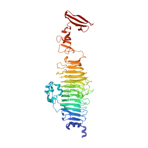
An essential serotype recognition pocket on phage P22 tailspike protein forces Salmonella enterica serovar Paratyphi A O-antigen fragments to bind as nonsolution conformers.
Andres, D., Gohlke, U., Broeker, N.K., Schulze, S., Rabsch, W., Heinemann, U., Barbirz, S., Seckler, R.(2013) Glycobiology 23: 486-494
- PubMed: 23292517
- DOI: https://doi.org/10.1093/glycob/cws224
- Primary Citation of Related Structures:
3TH0 - PubMed Abstract:
Bacteriophage P22 recognizes O-antigen polysaccharides of Salmonella enterica subsp. enterica (S.) with its tailspike protein (TSP). In the serovars S. Typhimurium, S. Enteritidis, and S. Paratyphi A, the tetrasaccharide repeat units of the respective O-antigens consist of an identical main chain trisaccharide but different 3,6-dideoxyhexose substituents. Here, the epimers abequose, tyvelose and paratose determine the specific serotype. P22 TSP recognizes O-antigen octasaccharides in an extended binding site with a single 3,6-dideoxyhexose binding pocket. We have isolated S. Paratyphi A octasaccharides which were not available previously and determined the crystal structure of their complex with P22 TSP. We discuss our data together with crystal structures of complexes with S. Typhimurium and S. Enteritidis octasaccharides determined earlier. Isothermal titration calorimetry showed that S. Paratyphi A octasaccharide binds P22 TSP less tightly, with a difference in binding free energy of ∼7 kJ mol(-1) at 20°C compared with S. Typhimurium and S. Enteritidis octasaccharides. Individual protein-carbohydrate contacts were probed by amino acid replacements showing that the dideoxyhexose pocket contributes to binding of all three serotypes. However, S. Paratyphi A octasaccharides bind in a conformation with an energetically unfavorable ϕ/ψ glycosidic bond angle combination. In contrast, octasaccharides from the other serotypes bind as solution-like conformers. Two water molecules are conserved in all P22 TSP complexes with octasaccharides of different serotypes. They line the dideoxyhexose binding pocket and force the S. Paratyphi A octasaccharides to bind as nonsolution conformers. This emphasizes the role of solvent as part of carbohydrate binding sites.
Organizational Affiliation:
Physikalische Biochemie, Universität Potsdam, Karl-Liebknecht-Str. 24-25, 14476 Potsdam, Germany.