3TB7

The type I crystal structure of Streptococcus agalactiae sortase C1


Experimental Data Snapshot

  • Method: X-RAY DIFFRACTION
  • Resolution: 2.45 Å
  • R-Value Free: 0.308 
  • R-Value Work: 0.228 
  • R-Value Observed: 0.232 

wwPDB Validation   3D Report Full Report


This is version 1.4 of the entry. See complete history


Literature

The Crystal Structure Analysis of Group B Streptococcus Sortase C1: A Model for the "Lid" Movement upon Substrate Binding.

Khare, B.Fu, Z.Q.Huang, I.H.Ton-That, H.Narayana, S.V.

(2011) J Mol Biol 414: 563-577

  • DOI: https://doi.org/10.1016/j.jmb.2011.10.017
  • Primary Citation of Related Structures:  
    3TB7, 3TBE

  • PubMed Abstract: 

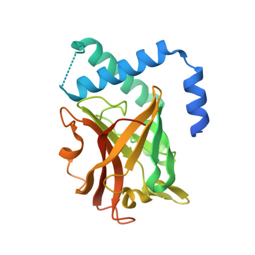
    A unique feature of the class-C-type sortases, enzymes essential for Gram-positive pilus biogenesis, is the presence of a flexible "lid" anchored in the active site. However, the mechanistic details of the "lid" displacement, suggested to be a critical prelude for enzyme catalysis, are not yet known. This is partly due to the absence of enzyme-substrate and enzyme-inhibitor complex crystal structures. We have recently described the crystal structures of the Streptococcus agalactiae SAG2603 V/R sortase SrtC1 in two space groups (type II and type III) and that of its "lid" mutant and proposed a role of the "lid" as a protector of the active-site hydrophobic environment. Here, we report the crystal structures of SAG2603 V/R sortase C1 in a different space group (type I) and that of its complex with a small-molecule cysteine protease inhibitor. We observe that the catalytic Cys residue is covalently linked to the small-molecule inhibitor without lid displacement. However, the type I structure provides a view of the sortase SrtC1 lid displacement while having structural elements similar to a substrate sorting motif suitably positioned in the active site. We propose that these major conformational changes seen in the presence of a substrate mimic in the active site may represent universal features of class C sortase substrate recognition and enzyme activation.


  • Organizational Affiliation

    Center for Biophysical Sciences and Engineering, University of Alabama at Birmingham, Birmingham, AL 35294, USA.


Macromolecules
Find similar proteins by:  (by identity cutoff)  |  3D Structure
Entity ID: 1
MoleculeChains Sequence LengthOrganismDetailsImage
Sortase family protein230Streptococcus agalactiae serogroup VMutation(s): 0 
Gene Names: SAG0647
EC: 3.4.22
UniProt
Find proteins for Q8E0S7 (Streptococcus agalactiae serotype V (strain ATCC BAA-611 / 2603 V/R))
Explore Q8E0S7 
Go to UniProtKB:  Q8E0S7
Entity Groups  
Sequence Clusters30% Identity50% Identity70% Identity90% Identity95% Identity100% Identity
UniProt GroupQ8E0S7
Sequence Annotations
Expand
  • Reference Sequence
Experimental Data & Validation

Experimental Data

  • Method: X-RAY DIFFRACTION
  • Resolution: 2.45 Å
  • R-Value Free: 0.308 
  • R-Value Work: 0.228 
  • R-Value Observed: 0.232 
  • Space Group: C 1 2 1
Unit Cell:
Length ( Å )Angle ( ˚ )
a = 78.59α = 90
b = 46.93β = 114.99
c = 66.57γ = 90
Software Package:
Software NamePurpose
REFMACrefinement
SGXPROmodel building
SOLVEphasing
CNSrefinement
HKL-2000data collection
HKL-2000data reduction
HKL-2000data scaling
SGXPROphasing

Structure Validation

View Full Validation Report


Entry History 

Deposition Data

  • Released Date: 2011-10-26 
  • Deposition Author(s): Khare, B.

Revision History  (Full details and data files)

  • Version 1.0: 2011-10-26
    Type: Initial release
  • Version 1.1: 2011-11-09
    Changes: Database references
  • Version 1.2: 2011-12-21
    Changes: Database references
  • Version 1.3: 2019-07-17
    Changes: Data collection, Refinement description
  • Version 1.4: 2024-02-28
    Changes: Data collection, Database references