3T0F

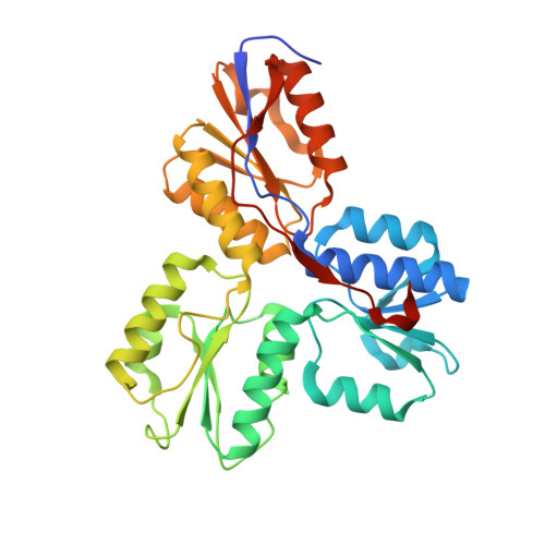
IspH:HMBPP (substrate) structure of the E126D mutant


Experimental Data Snapshot

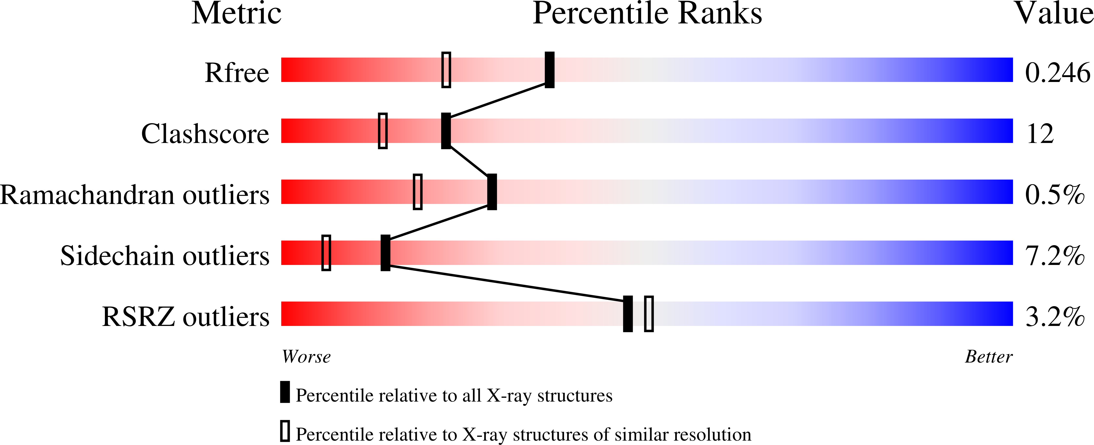
  • Method: X-RAY DIFFRACTION
  • Resolution: 1.90 Å
  • R-Value Free: 0.242 
  • R-Value Work: 0.201 
  • R-Value Observed: 0.203 

wwPDB Validation   3D Report Full Report


This is version 1.3 of the entry. See complete history


Literature

Crystal Structures of Mutant IspH Proteins Reveal a Rotation of the Substrate's Hydroxymethyl Group during Catalysis.

Span, I.Grawert, T.Bacher, A.Eisenreich, W.Groll, M.

(2012) J Mol Biol 416: 1-9

  • DOI: https://doi.org/10.1016/j.jmb.2011.11.033
  • Primary Citation of Related Structures:  
    3SZL, 3SZO, 3SZU, 3T0F, 3T0G

  • PubMed Abstract: 

    Isoprenoids derive from two universal precursors, isopentenyl diphosphate and dimethylallyl diphosphate, which in most human pathogens are synthesized in the deoxyxylulose phosphate pathway. The last step of this pathway is the conversion of (E)-1-hydroxy-2-methylbut-2-enyl-4-diphosphate into a mixture of isopentenyl diphosphate and dimethylallyl diphosphate catalyzed by the iron-sulfur protein IspH. The crystal structures reported here of the IspH mutant proteins T167C, E126D and E126Q reveal an alternative substrate conformation compared to the wild-type structure. Thus, the previously observed alkoxide complex decomposes, and the substrate's hydroxymethyl group rotates to interact with Glu126. The carboxyl group of Glu126 then donates a proton to the hydroxyl group to enable water elimination. The structural and functional studies provide further knowledge of the IspH reaction mechanism, which opens up new routes to inhibitor design against malaria and tuberculosis.


  • Organizational Affiliation

    Chemistry Department, Center for Integrated Protein Science, Chair of Biochemistry, Technische Universität München, Lichtenbergstrasse 4, 85747 Garching, Germany.


Macromolecules
Find similar proteins by:  (by identity cutoff)  |  3D Structure
Entity ID: 1
MoleculeChains Sequence LengthOrganismDetailsImage
4-hydroxy-3-methylbut-2-enyl diphosphate reductase
A, B
328Escherichia coli K-12Mutation(s): 1 
Gene Names: ispHlytByaaEb0029JW0027
EC: 1.17.1.2
UniProt
Find proteins for P62623 (Escherichia coli (strain K12))
Explore P62623 
Go to UniProtKB:  P62623
Entity Groups  
Sequence Clusters30% Identity50% Identity70% Identity90% Identity95% Identity100% Identity
UniProt GroupP62623
Sequence Annotations
Expand
  • Reference Sequence
Experimental Data & Validation

Experimental Data

  • Method: X-RAY DIFFRACTION
  • Resolution: 1.90 Å
  • R-Value Free: 0.242 
  • R-Value Work: 0.201 
  • R-Value Observed: 0.203 
  • Space Group: P 21 21 21
Unit Cell:
Length ( Å )Angle ( ˚ )
a = 64.85α = 90
b = 80.18β = 90
c = 114.51γ = 90
Software Package:
Software NamePurpose
XDSdata scaling
REFMACrefinement
XDSdata reduction
XSCALEdata scaling
REFMACphasing

Structure Validation

View Full Validation Report


Entry History 

Deposition Data

Revision History  (Full details and data files)

  • Version 1.0: 2011-11-30
    Type: Initial release
  • Version 1.1: 2012-01-11
    Changes: Database references
  • Version 1.2: 2012-02-15
    Changes: Database references
  • Version 1.3: 2023-09-13
    Changes: Data collection, Database references, Derived calculations, Refinement description