3SQG

Crystal structure of a methyl-coenzyme M reductase purified from Black Sea mats


Experimental Data Snapshot

  • Method: X-RAY DIFFRACTION
  • Resolution: 2.10 Å
  • R-Value Free: 0.206 
  • R-Value Work: 0.161 
  • R-Value Observed: 0.163 

wwPDB Validation   3D Report Full Report


Ligand Structure Quality Assessment 


This is version 1.5 of the entry. See complete history


Literature

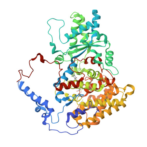
Structure of a methyl-coenzyme M reductase from Black Sea mats that oxidize methane anaerobically.

Shima, S.Krueger, M.Weinert, T.Demmer, U.Kahnt, J.Thauer, R.K.Ermler, U.

(2011) Nature 481: 98-101

  • DOI: https://doi.org/10.1038/nature10663
  • Primary Citation of Related Structures:  
    3SQG

  • PubMed Abstract: 

    The anaerobic oxidation of methane (AOM) with sulphate, an area currently generating great interest in microbiology, is accomplished by consortia of methanotrophic archaea (ANME) and sulphate-reducing bacteria. The enzyme activating methane in methanotrophic archaea has tentatively been identified as a homologue of methyl-coenzyme M reductase (MCR) that catalyses the methane-forming step in methanogenic archaea. Here we report an X-ray structure of the 280 kDa heterohexameric ANME-1 MCR complex. It was crystallized uniquely from a protein ensemble purified from consortia of microorganisms collected with a submersible from a Black Sea mat catalysing AOM with sulphate. Crystals grown from the heterogeneous sample diffract to 2.1 Å resolution and consist of a single ANME-1 MCR population, demonstrating the strong selective power of crystallization. The structure revealed ANME-1 MCR in complex with coenzyme M and coenzyme B, indicating the same substrates for MCR from methanotrophic and methanogenic archaea. Differences between the highly similar structures of ANME-1 MCR and methanogenic MCR include a F(430) modification, a cysteine-rich patch and an altered post-translational amino acid modification pattern, which may tune the enzymes for their functions in different biological contexts.


  • Organizational Affiliation

    Max Planck Institute for Terrestrial Microbiology, Karl-Frisch-Strasse 10, D-35043 Marburg, Germany. shima@mpi-marburg.mpg.de


Macromolecules
Find similar proteins by:  (by identity cutoff)  |  3D Structure
Entity ID: 1
MoleculeChains Sequence LengthOrganismDetailsImage
Methyl coenzyme M reductase, alpha subunit
A, D, G
579uncultured archaeonMutation(s): 0 
EC: 2.8.4.1
UniProt
Find proteins for D1JBK4 (Uncultured archaeon)
Explore D1JBK4 
Go to UniProtKB:  D1JBK4
Entity Groups  
Sequence Clusters30% Identity50% Identity70% Identity90% Identity95% Identity100% Identity
UniProt GroupD1JBK4
Sequence Annotations
Expand
  • Reference Sequence
Find similar proteins by:  (by identity cutoff)  |  3D Structure
Entity ID: 2
MoleculeChains Sequence LengthOrganismDetailsImage
Methyl-coenzyme M reductase, beta subunit
B, E, H
433uncultured archaeonMutation(s): 0 
EC: 2.8.4.1
UniProt
Find proteins for D1JBK2 (Uncultured archaeon)
Explore D1JBK2 
Go to UniProtKB:  D1JBK2
Entity Groups  
Sequence Clusters30% Identity50% Identity70% Identity90% Identity95% Identity100% Identity
UniProt GroupD1JBK2
Sequence Annotations
Expand
  • Reference Sequence
Find similar proteins by:  (by identity cutoff)  |  3D Structure
Entity ID: 3
MoleculeChains Sequence LengthOrganismDetailsImage
Methyl-coenzyme M reductase, gamma subunit
C, F, I
279uncultured archaeonMutation(s): 0 
EC: 2.8.4.1
UniProt
Find proteins for D1JBK3 (Uncultured archaeon)
Explore D1JBK3 
Go to UniProtKB:  D1JBK3
Entity Groups  
Sequence Clusters30% Identity50% Identity70% Identity90% Identity95% Identity100% Identity
UniProt GroupD1JBK3
Sequence Annotations
Expand
  • Reference Sequence
Small Molecules
Ligands 10 Unique
IDChains Name / Formula / InChI Key2D Diagram3D Interactions
M43
Query on M43

Download Ideal Coordinates CCD File 
JA [auth G],
L [auth A],
Z [auth D]
(17[2]S)-17[2]-methylthio-coenzyme F43
C43 H53 N6 Ni O13 S
UOYNURPVGLQPFO-AEEVBHTRSA-M
TP7
Query on TP7

Download Ideal Coordinates CCD File 
J [auth A],
KA [auth G],
M [auth A]
Coenzyme B
C11 H22 N O7 P S
JBJSVEVEEGOEBZ-SCZZXKLOSA-N
P6G
Query on P6G

Download Ideal Coordinates CCD File 
U [auth B]HEXAETHYLENE GLYCOL
C12 H26 O7
IIRDTKBZINWQAW-UHFFFAOYSA-N
1PE
Query on 1PE

Download Ideal Coordinates CCD File 
BA [auth D]
GA [auth E]
IA [auth F]
MA [auth G]
N [auth A]
BA [auth D],
GA [auth E],
IA [auth F],
MA [auth G],
N [auth A],
QA [auth H]
PENTAETHYLENE GLYCOL
C10 H22 O6
JLFNLZLINWHATN-UHFFFAOYSA-N
PGE
Query on PGE

Download Ideal Coordinates CCD File 
CA [auth D]
DA [auth D]
NA [auth G]
O [auth A]
OA [auth G]
CA [auth D],
DA [auth D],
NA [auth G],
O [auth A],
OA [auth G],
P [auth A]
TRIETHYLENE GLYCOL
C6 H14 O4
ZIBGPFATKBEMQZ-UHFFFAOYSA-N
COM
Query on COM

Download Ideal Coordinates CCD File 
AA [auth D],
K [auth A],
LA [auth G]
1-THIOETHANESULFONIC ACID
C2 H6 O3 S2
ZNEWHQLOPFWXOF-UHFFFAOYSA-N
SO4
Query on SO4

Download Ideal Coordinates CCD File 
EA [auth D]
FA [auth D]
Q [auth A]
R [auth A]
RA [auth H]
EA [auth D],
FA [auth D],
Q [auth A],
R [auth A],
RA [auth H],
SA [auth I],
V [auth B],
W [auth B],
Y [auth C]
SULFATE ION
O4 S
QAOWNCQODCNURD-UHFFFAOYSA-L
GOL
Query on GOL

Download Ideal Coordinates CCD File 
HA [auth E],
X [auth C]
GLYCEROL
C3 H8 O3
PEDCQBHIVMGVHV-UHFFFAOYSA-N
CA
Query on CA

Download Ideal Coordinates CCD File 
PA [auth G],
T [auth A]
CALCIUM ION
Ca
BHPQYMZQTOCNFJ-UHFFFAOYSA-N
CL
Query on CL

Download Ideal Coordinates CCD File 
S [auth A]CHLORIDE ION
Cl
VEXZGXHMUGYJMC-UHFFFAOYSA-M
Modified Residues  4 Unique
IDChains TypeFormula2D DiagramParent
0AF
Query on 0AF
A, D, G
L-PEPTIDE LINKINGC11 H12 N2 O3TRP
GL3
Query on GL3
A, D, G
L-PEPTIDE LINKINGC2 H5 N O SGLY
MHO
Query on MHO
A, D, G
L-PEPTIDE LINKINGC5 H11 N O3 SMET
MHS
Query on MHS
A, D, G
L-PEPTIDE LINKINGC7 H11 N3 O2HIS
Experimental Data & Validation

Experimental Data

  • Method: X-RAY DIFFRACTION
  • Resolution: 2.10 Å
  • R-Value Free: 0.206 
  • R-Value Work: 0.161 
  • R-Value Observed: 0.163 
  • Space Group: C 2 2 21
Unit Cell:
Length ( Å )Angle ( ˚ )
a = 128.86α = 90
b = 412.49β = 90
c = 165.51γ = 90
Software Package:
Software NamePurpose
PHASERphasing
PHENIXrefinement
XDSdata reduction
XSCALEdata scaling

Structure Validation

View Full Validation Report


Ligand Structure Quality Assessment 


Entry History 

Deposition Data

Revision History  (Full details and data files)

  • Version 1.0: 2011-11-30
    Type: Initial release
  • Version 1.1: 2011-12-28
    Changes: Database references
  • Version 1.2: 2012-01-04
    Changes: Database references
  • Version 1.3: 2015-05-20
    Changes: Non-polymer description
  • Version 1.4: 2015-05-27
    Changes: Other
  • Version 1.5: 2017-11-08
    Changes: Refinement description