3SI0

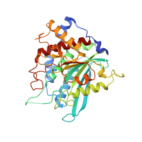
Structure of glycosylated human glutaminyl cyclase


Experimental Data Snapshot

  • Method: X-RAY DIFFRACTION
  • Resolution: 2.10 Å
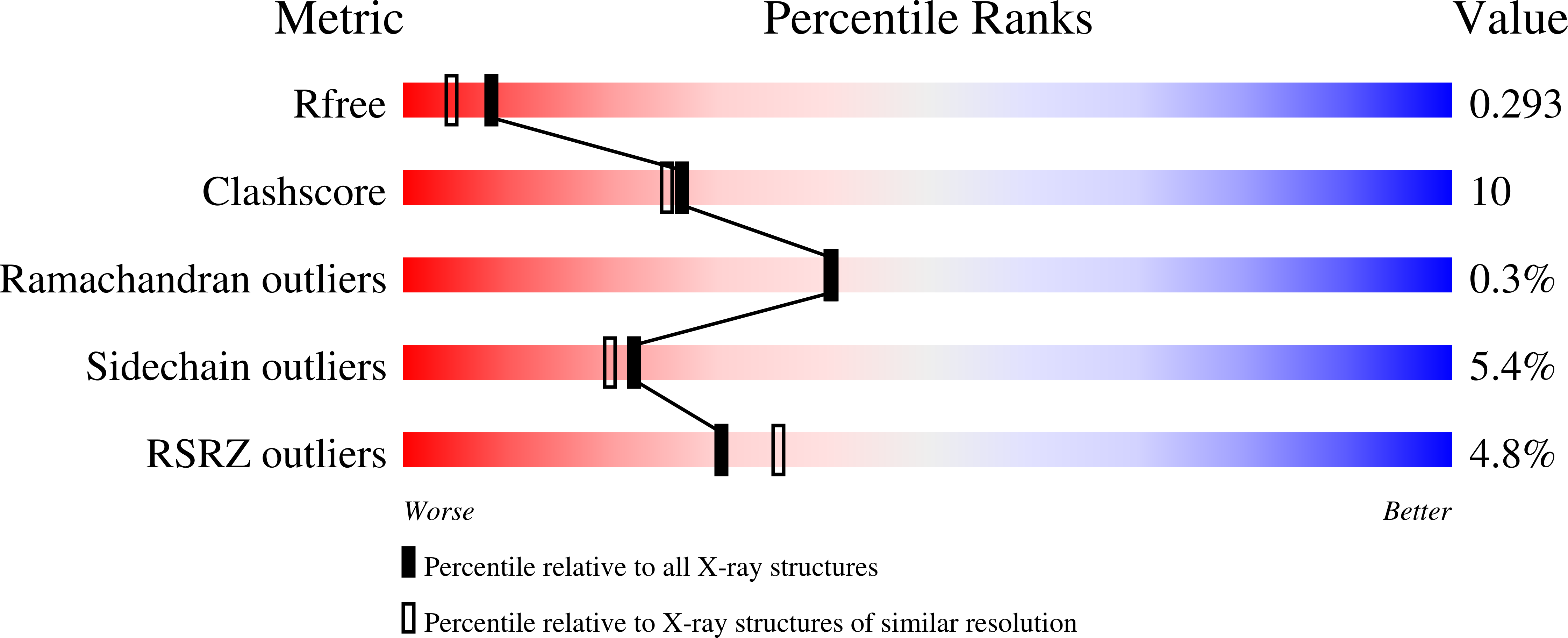
  • R-Value Free: 0.264 
  • R-Value Work: 0.204 
  • R-Value Observed: 0.207 

wwPDB Validation   3D Report Full Report


This is version 2.1 of the entry. See complete history


Literature

Structures of Glycosylated Mammalian Glutaminyl Cyclases Reveal Conformational Variability near the Active Center.

Ruiz-Carrillo, D.Koch, B.Parthier, C.Wermann, M.Dambe, T.Buchholz, M.Ludwig, H.H.Heiser, U.Rahfeld, J.U.Stubbs, M.T.Schilling, S.Demuth, H.U.

(2011) Biochemistry 50: 6280-6288

  • DOI: https://doi.org/10.1021/bi200249h
  • Primary Citation of Related Structures:  
    3SI0, 3SI1, 3SI2

  • PubMed Abstract: 

    Formation of N-terminal pyroglutamate (pGlu or pE) from glutaminyl or glutamyl precursors is catalyzed by glutaminyl cyclases (QC). As the formation of pGlu-amyloid has been linked with Alzheimer's disease, inhibitors of QCs are currently the subject of intense development. Here, we report three crystal structures of N-glycosylated mammalian QC from humans (hQC) and mice (mQC). Whereas the overall structures of the enzymes are similar to those reported previously, two surface loops in the neighborhood of the active center exhibit conformational variability. Furthermore, two conserved cysteine residues form a disulfide bond at the base of the active center that was not present in previous reports of hQC structure. Site-directed mutagenesis suggests a structure-stabilizing role of the disulfide bond. At the entrance to the active center, the conserved tryptophan residue, W(207), which displayed multiple orientations in previous structure, shows a single conformation in both glycosylated human and murine QCs. Although mutagenesis of W(207) into leucine or glutamine altered substrate conversion significantly, the binding constants of inhibitors such as the highly potent PQ50 (PBD150) were minimally affected. The crystal structure of PQ50 bound to the active center of murine QC reveals principal binding determinants provided by the catalytic zinc ion and a hydrophobic funnel. This study presents a first comparison of two mammalian QCs containing typical, conserved post-translational modifications.


  • Organizational Affiliation

    Probiodrug AG, Weinbergweg 22, D-06120 Halle, Saale, Germany.


Macromolecules
Find similar proteins by:  (by identity cutoff)  |  3D Structure
Entity ID: 1
MoleculeChains Sequence LengthOrganismDetailsImage
Glutaminyl-peptide cyclotransferase330Homo sapiensMutation(s): 0 
Gene Names: QPCT
EC: 2.3.2.5
UniProt & NIH Common Fund Data Resources
Find proteins for Q16769 (Homo sapiens)
Explore Q16769 
Go to UniProtKB:  Q16769
PHAROS:  Q16769
GTEx:  ENSG00000115828 
Entity Groups  
Sequence Clusters30% Identity50% Identity70% Identity90% Identity95% Identity100% Identity
UniProt GroupQ16769
Sequence Annotations
Expand
  • Reference Sequence
Oligosaccharides

Help

Entity ID: 2
MoleculeChains Length2D Diagram Glycosylation3D Interactions
2-acetamido-2-deoxy-beta-D-glucopyranose-(1-4)-2-acetamido-2-deoxy-beta-D-glucopyranose
B
2N-Glycosylation
Glycosylation Resources
GlyTouCan:  G42666HT
GlyCosmos:  G42666HT
GlyGen:  G42666HT
Experimental Data & Validation

Experimental Data

  • Method: X-RAY DIFFRACTION
  • Resolution: 2.10 Å
  • R-Value Free: 0.264 
  • R-Value Work: 0.204 
  • R-Value Observed: 0.207 
  • Space Group: C 1 2 1
Unit Cell:
Length ( Å )Angle ( ˚ )
a = 82.408α = 90
b = 63.688β = 105.76
c = 77.159γ = 90
Software Package:
Software NamePurpose
SCALAdata scaling
PHASERphasing
REFMACrefinement
PDB_EXTRACTdata extraction
MAR345dtbdata collection
XSCALEdata scaling

Structure Validation

View Full Validation Report


Entry History 

Deposition Data

Revision History  (Full details and data files)

  • Version 1.0: 2011-06-29
    Type: Initial release
  • Version 1.1: 2011-07-13
    Changes: Version format compliance
  • Version 1.2: 2011-07-27
    Changes: Database references
  • Version 1.3: 2011-08-31
    Changes: Structure summary
  • Version 1.4: 2017-11-08
    Changes: Refinement description
  • Version 2.0: 2020-07-29
    Type: Remediation
    Reason: Carbohydrate remediation
    Changes: Atomic model, Data collection, Database references, Derived calculations, Structure summary
  • Version 2.1: 2023-09-13
    Changes: Data collection, Database references, Refinement description, Structure summary