3S6T

Crystal Structure of insect beta-N-acetyl-D-hexosaminidase OfHex1 V327G complexed with PUGNAc


Experimental Data Snapshot

  • Method: X-RAY DIFFRACTION
  • Resolution: 2.30 Å
  • R-Value Free: 0.227 
  • R-Value Work: 0.210 
  • R-Value Observed: 0.211 

wwPDB Validation   3D Report Full Report


Ligand Structure Quality Assessment 


This is version 1.1 of the entry. See complete history


Literature

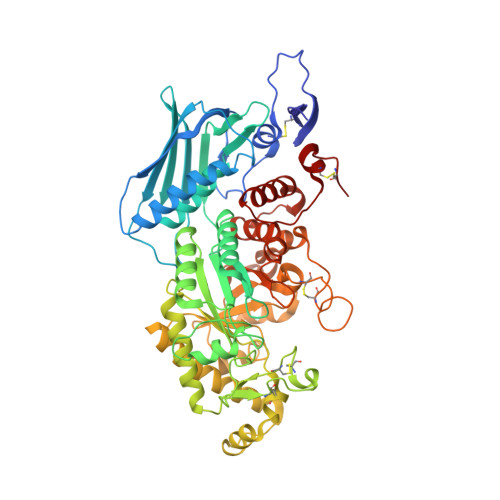
Active-pocket size differentiating insectile from bacterial chitinolytic beta-N-acetyl-D-hexosaminidases.

Liu, T.Zhang, H.Liu, F.Chen, L.Shen, X.Yang, Q.

(2011) Biochem J 438: 467-474

  • DOI: https://doi.org/10.1042/BJ20110390
  • Primary Citation of Related Structures:  
    3OZP, 3S6T

  • PubMed Abstract: 

    Chitinolytic β-N-acetyl-D-hexosaminidase is a branch of the GH20 (glycoside hydrolase family 20) β-N-acetyl-D-hexosaminidases that is only distributed in insects and micro-organisms, and is therefore a potential target for the action of insecticides. PUGNAc [O-(2-acetamido-2-deoxy-D-glucopyransylidene)-amino-N-phenylcarbamate] was initially identified as an inhibitor against GH20 β-N-acetyl-D-hexosaminidases. So far no crystal structure of PUGNAc in complex with any GH20 β-N-acetyl-D-hexosaminidase has been reported. We show in the present study that the sensitivities of chitinolytic β-N-acetyl-D-hexosaminidases towards PUGNAc can vary by 100-fold, with the order being OfHex1 (Ostrinia furnacalis β-N-acetyl-D-hexosaminidase)


  • Organizational Affiliation

    School of Life Science and Biotechnology, Dalian University of Technology, Dalian 116024, China.


Macromolecules
Find similar proteins by:  (by identity cutoff)  |  3D Structure
Entity ID: 1
MoleculeChains Sequence LengthOrganismDetailsImage
N-acetylglucosaminidase575Ostrinia furnacalisMutation(s): 1 
EC: 3.2.1.52
UniProt
Find proteins for Q06GJ0 (Ostrinia furnacalis)
Explore Q06GJ0 
Go to UniProtKB:  Q06GJ0
Entity Groups  
Sequence Clusters30% Identity50% Identity70% Identity90% Identity95% Identity100% Identity
UniProt GroupQ06GJ0
Sequence Annotations
Expand
  • Reference Sequence
Small Molecules
Ligands 1 Unique
IDChains Name / Formula / InChI Key2D Diagram3D Interactions
OAN
Query on OAN

Download Ideal Coordinates CCD File 
B [auth A]O-(2-ACETAMIDO-2-DEOXY D-GLUCOPYRANOSYLIDENE) AMINO-N-PHENYLCARBAMATE
C15 H19 N3 O7
PBLNJFVQMUMOJY-JXZOILRNSA-N
Binding Affinity Annotations 
IDSourceBinding Affinity
OAN PDBBind:  3S6T Ki: 45 (nM) from 1 assay(s)
Binding MOAD:  3S6T Ki: 45 (nM) from 1 assay(s)
Experimental Data & Validation

Experimental Data

  • Method: X-RAY DIFFRACTION
  • Resolution: 2.30 Å
  • R-Value Free: 0.227 
  • R-Value Work: 0.210 
  • R-Value Observed: 0.211 
  • Space Group: P 32 2 1
Unit Cell:
Length ( Å )Angle ( ˚ )
a = 108.79α = 90
b = 108.79β = 90
c = 175.05γ = 120
Software Package:
Software NamePurpose
CrystalCleardata collection
AMoREphasing
REFMACrefinement
CrystalCleardata reduction
CrystalCleardata scaling

Structure Validation

View Full Validation Report


Ligand Structure Quality Assessment 


Entry History 

Deposition Data

Revision History  (Full details and data files)

  • Version 1.0: 2012-04-11
    Type: Initial release
  • Version 1.1: 2023-11-01
    Changes: Data collection, Database references, Derived calculations, Refinement description