


Crystal structure of a Hypothetical Glycosyl hydrolase (BDI_2473) from Parabacteroides distasonis ATCC 8503 at 1.85 A resolution
Joint Center for Structural Genomics (JCSG)To be published.
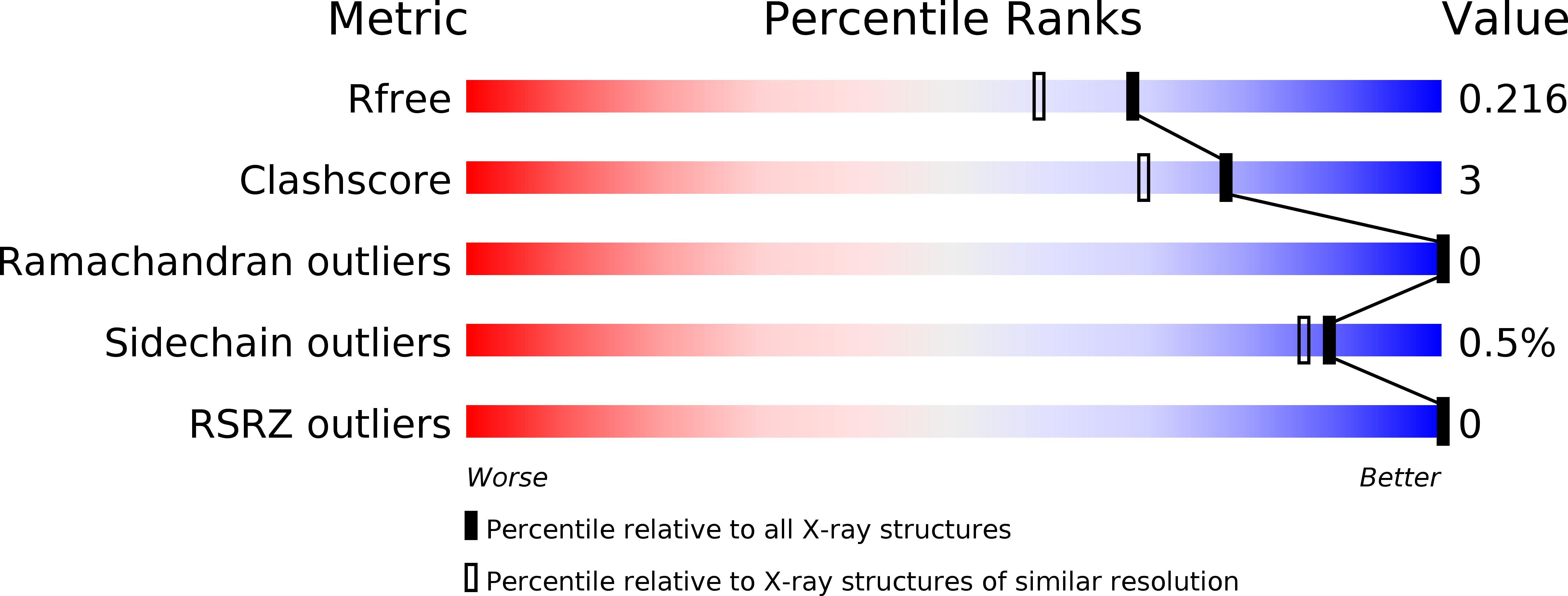
Experimental Data Snapshot
wwPDB Validation 3D Report Full Report
Entity ID: 1 | |||||
|---|---|---|---|---|---|
| Molecule | Chains | Sequence Length | Organism | Details | Image |
| Putative glycosylhydrolase | 214 | Parabacteroides distasonis ATCC 8503 | Mutation(s): 0 Gene Names: BDI_2473 | 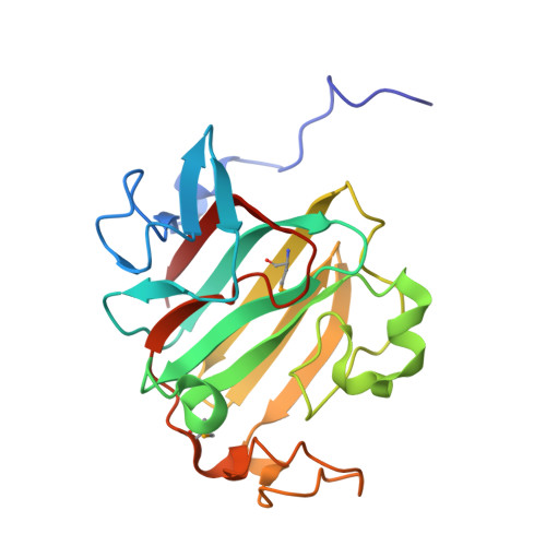 | |
UniProt | |||||
Find proteins for A6LES9 (Parabacteroides distasonis (strain ATCC 8503 / DSM 20701 / CIP 104284 / JCM 5825 / NCTC 11152)) Explore A6LES9 Go to UniProtKB: A6LES9 | |||||
Entity Groups | |||||
| Sequence Clusters | 30% Identity50% Identity70% Identity90% Identity95% Identity100% Identity | ||||
| UniProt Group | A6LES9 | ||||
Sequence AnnotationsExpand | |||||
| |||||
| Ligands 2 Unique | |||||
|---|---|---|---|---|---|
| ID | Chains | Name / Formula / InChI Key | 2D Diagram | 3D Interactions | |
| IOD Query on IOD | B [auth A], C [auth A], D [auth A] | IODIDE ION I XMBWDFGMSWQBCA-UHFFFAOYSA-M |  | ||
| EDO Query on EDO | E [auth A] F [auth A] G [auth A] H [auth A] I [auth A] | 1,2-ETHANEDIOL C2 H6 O2 LYCAIKOWRPUZTN-UHFFFAOYSA-N |  | ||
| Modified Residues 1 Unique | |||||
|---|---|---|---|---|---|
| ID | Chains | Type | Formula | 2D Diagram | Parent |
| MSE Query on MSE | A | L-PEPTIDE LINKING | C5 H11 N O2 Se |  | MET |
| Length ( Å ) | Angle ( ˚ ) |
|---|---|
| a = 41.062 | α = 90 |
| b = 60.473 | β = 90 |
| c = 77.221 | γ = 90 |
| Software Name | Purpose |
|---|---|
| MolProbity | model building |
| PDB_EXTRACT | data extraction |
| SHELX | phasing |
| SHARP | phasing |
| XSCALE | data scaling |
| REFMAC | refinement |
| XDS | data reduction |
| SHELXD | phasing |
| autoSHARP | phasing |
RCSB PDB (citation) is hosted by
RCSB PDB is a member of the

