


Cellobiose phosphorylase: reconstructing the structural itinerary along the catalytic pathway
Van Hoorebeke, A., Stout, J., Soetaert, W., Van Beeumen, J., Desmet, T., Savvides, S.To be published.
Experimental Data Snapshot
Entity ID: 1 | |||||
|---|---|---|---|---|---|
| Molecule | Chains | Sequence Length | Organism | Details | Image |
| Cellobiose phosphorylase | 822 | Cellulomonas uda | Mutation(s): 0 EC: 2.4.1.20 | 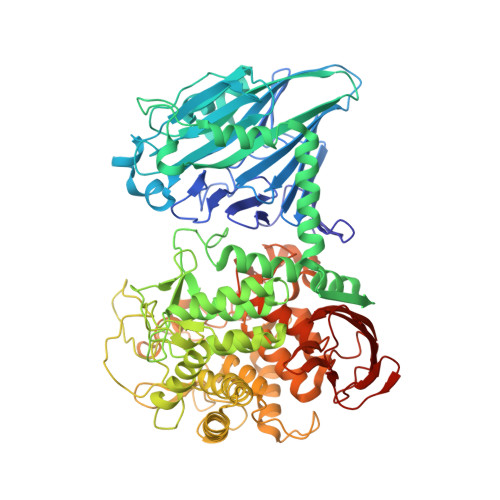 | |
UniProt | |||||
Find proteins for Q7WTR6 (Cellulomonas uda) Explore Q7WTR6 Go to UniProtKB: Q7WTR6 | |||||
Entity Groups | |||||
| Sequence Clusters | 30% Identity50% Identity70% Identity90% Identity95% Identity100% Identity | ||||
| UniProt Group | Q7WTR6 | ||||
Sequence AnnotationsExpand | |||||
| |||||
| Ligands 1 Unique | |||||
|---|---|---|---|---|---|
| ID | Chains | Name / Formula / InChI Key | 2D Diagram | 3D Interactions | |
| GLC Query on GLC | C [auth A], D [auth A], E [auth B], F [auth B] | alpha-D-glucopyranose C6 H12 O6 WQZGKKKJIJFFOK-DVKNGEFBSA-N |  | ||
| Length ( Å ) | Angle ( ˚ ) |
|---|---|
| a = 85.417 | α = 90 |
| b = 102.74 | β = 96.44 |
| c = 98.305 | γ = 90 |
| Software Name | Purpose |
|---|---|
| PHENIX | refinement |
| PDB_EXTRACT | data extraction |
| MOSFLM | data reduction |
| SCALA | data scaling |
| PHASER | phasing |
RCSB PDB (citation) is hosted by
RCSB PDB is a member of the

