3RTY

Structure of an Enclosed Dimer Formed by The Drosophila Period Protein


Experimental Data Snapshot

  • Method: X-RAY DIFFRACTION
  • Resolution: 2.85 Å
  • R-Value Free: 0.289 
  • R-Value Work: 0.239 
  • R-Value Observed: 0.239 

wwPDB Validation   3D Report Full Report


This is version 1.0 of the entry. See complete history


Literature

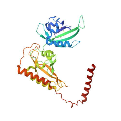
Structure of an enclosed dimer formed by the Drosophila period protein.

King, H.A.Hoelz, A.Crane, B.R.Young, M.W.

(2011) J Mol Biol 413: 561-572

  • DOI: https://doi.org/10.1016/j.jmb.2011.08.048
  • Primary Citation of Related Structures:  
    3RTY

  • PubMed Abstract: 

    Period (PER) is the major transcription inhibitor in metazoan circadian clocks and lies at the center of several feedback loops that regulate gene expression. Dimerization of Drosophila PER influences nuclear translocation, repressor activity, and behavioral rhythms. The structure of a central, 346-residue PER fragment reveals two associated PAS (Per-Arnt-Sim) domains followed by a protruding α-helical extension (αF). A closed, pseudo-symmetric dimer forms from a cross handshake interaction of the N-terminal PAS domain with αF of the opposing subunit. Strikingly, a shift of αF against the PAS β-sheet generates two alternative subunit interfaces in the dimer. Taken together with a previously reported PER structure in which αF extends, these data indicate that αF unlatches to switch association of PER with itself to its partner Timeless. The variable positions of the αF helix provide snapshots of a helix dissociation mechanism that has relevance to other PAS protein systems. Conservation of PER interaction residues among a family of PAS-AB-containing transcription factors suggests that contacts mediating closed PAS-AB dimers serve a general function.


  • Organizational Affiliation

    Laboratory of Genetics, The Rockefeller University, New York, NY 10065, USA.


Macromolecules
Find similar proteins by:  (by identity cutoff)  |  3D Structure
Entity ID: 1
MoleculeChains Sequence LengthOrganismDetailsImage
Period circadian protein
A, B, C, D, E
A, B, C, D, E, F, G, H
339Drosophila melanogasterMutation(s): 0 
Gene Names: perCG2647
UniProt
Find proteins for P07663 (Drosophila melanogaster)
Explore P07663 
Go to UniProtKB:  P07663
Entity Groups  
Sequence Clusters30% Identity50% Identity70% Identity90% Identity95% Identity100% Identity
UniProt GroupP07663
Sequence Annotations
Expand
  • Reference Sequence
Small Molecules
Ligands 1 Unique
IDChains Name / Formula / InChI Key2D Diagram3D Interactions
DTT
Query on DTT

Download Ideal Coordinates CCD File 
AA [auth F]
BA [auth F]
CA [auth F]
DA [auth G]
EA [auth G]
AA [auth F],
BA [auth F],
CA [auth F],
DA [auth G],
EA [auth G],
FA [auth G],
GA [auth H],
HA [auth H],
I [auth A],
IA [auth H],
J [auth A],
JA [auth H],
K [auth A],
L [auth A],
M [auth B],
N [auth B],
O [auth B],
P [auth B],
Q [auth C],
R [auth C],
S [auth C],
T [auth C],
U [auth D],
V [auth D],
W [auth D],
X [auth E],
Y [auth E],
Z [auth F]
2,3-DIHYDROXY-1,4-DITHIOBUTANE
C4 H10 O2 S2
VHJLVAABSRFDPM-IMJSIDKUSA-N
Experimental Data & Validation

Experimental Data

  • Method: X-RAY DIFFRACTION
  • Resolution: 2.85 Å
  • R-Value Free: 0.289 
  • R-Value Work: 0.239 
  • R-Value Observed: 0.239 
  • Space Group: P 1
Unit Cell:
Length ( Å )Angle ( ˚ )
a = 60.414α = 88.19
b = 94.697β = 89.63
c = 141.03γ = 89.87
Software Package:
Software NamePurpose
HKL-2000data collection
AMoREphasing
CNSrefinement
HKL-2000data reduction
HKL-2000data scaling

Structure Validation

View Full Validation Report


Entry History 

Deposition Data

Revision History  (Full details and data files)

  • Version 1.0: 2011-12-21
    Type: Initial release