3RS6

Crystal structure Dioclea virgata lectin in complexed with X-mannose


Experimental Data Snapshot

  • Method: X-RAY DIFFRACTION
  • Resolution: 1.80 Å
  • R-Value Free: 0.232 
  • R-Value Work: 0.189 
  • R-Value Observed: 0.192 

wwPDB Validation   3D Report Full Report


Ligand Structure Quality Assessment 


This is version 1.4 of the entry. See complete history


Literature

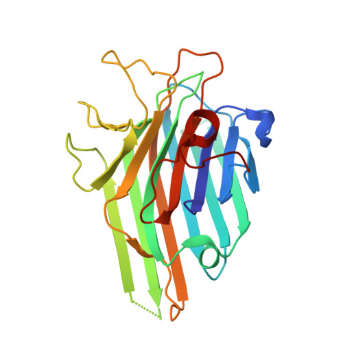
Structure of Dioclea virgata lectin: Relations between carbohydrate binding site and nitric oxide production.

Batista da Nobrega, R.Rocha, B.A.Gadelha, C.A.Santi-Gadelha, T.Pires, A.F.Assreuy, A.M.Nascimento, K.S.Nagano, C.S.Sampaio, A.H.Cavada, B.S.Delatorre, P.

(2012) Biochimie 94: 900-906

  • DOI: https://doi.org/10.1016/j.biochi.2011.12.009
  • Primary Citation of Related Structures:  
    3RRD, 3RS6

  • PubMed Abstract: 

    The lectin of Dioclea virgata (DvirL), both native and complexed with X-man, was submitted to X-ray diffraction analysis and the crystal structure was compared to that of other Diocleinae lectins in order to better understand differences in biological properties, especially with regard to the ability of lectins to induce nitric oxide (NO) production. An association was observed between the volume of the carbohydrate recognition domain (CRD), the ability to induce NO production and the relative positions of Tyr12, Arg228 and Leu99. Thus, differences in biological activity induced by Diocleinae lectins are related to the configuration of amino acid residues in the carbohydrate binding site and to the structural conformation of subsequent regions capable of influencing site-ligand interactions. In conclusion, the ability of Diocleinae lectins to induce NO production depends on CRD configuration.


  • Organizational Affiliation

    Departamento de Biologia Molecular, Universidade Federal da Paraíba, campus l, s/n, 58059-900 João Pessoa, Paraíba, Brazil.


Macromolecules
Find similar proteins by:  (by identity cutoff)  |  3D Structure
Entity ID: 1
MoleculeChains Sequence LengthOrganismDetailsImage
Lectin alpha chain237Dioclea virgataMutation(s): 0 
UniProt
Find proteins for P58907 (Dioclea virgata)
Explore P58907 
Go to UniProtKB:  P58907
Entity Groups  
Sequence Clusters30% Identity50% Identity70% Identity90% Identity95% Identity100% Identity
UniProt GroupP58907
Sequence Annotations
Expand
  • Reference Sequence
Small Molecules
Experimental Data & Validation

Experimental Data

  • Method: X-RAY DIFFRACTION
  • Resolution: 1.80 Å
  • R-Value Free: 0.232 
  • R-Value Work: 0.189 
  • R-Value Observed: 0.192 
  • Space Group: I 2 2 2
Unit Cell:
Length ( Å )Angle ( ˚ )
a = 64.55α = 90
b = 86.69β = 90
c = 90.28γ = 90
Software Package:
Software NamePurpose
MAR345dtbdata collection
MOLREPphasing
REFMACrefinement
MOSFLMdata reduction
SCALAdata scaling

Structure Validation

View Full Validation Report


Ligand Structure Quality Assessment 


Entry History 

Deposition Data

Revision History  (Full details and data files)

  • Version 1.0: 2012-02-08
    Type: Initial release
  • Version 1.1: 2012-02-22
    Changes: Database references
  • Version 1.2: 2018-01-24
    Changes: Structure summary
  • Version 1.3: 2020-07-29
    Type: Remediation
    Reason: Carbohydrate remediation
    Changes: Derived calculations, Structure summary
  • Version 1.4: 2024-02-28
    Changes: Data collection, Database references, Structure summary