3RFZ

Crystal structure of the FimD usher bound to its cognate FimC:FimH substrate


Experimental Data Snapshot

  • Method: X-RAY DIFFRACTION
  • Resolution: 2.80 Å
  • R-Value Free: 0.282 
  • R-Value Work: 0.223 
  • R-Value Observed: 0.226 

wwPDB Validation   3D Report Full Report


This is version 1.2 of the entry. See complete history


Literature

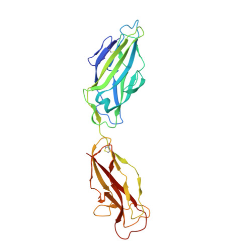
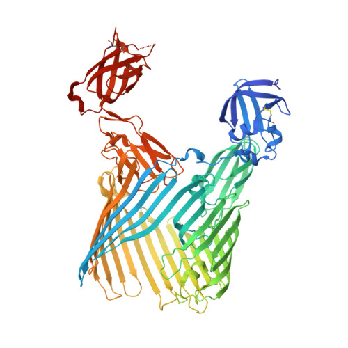
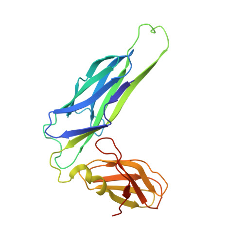
Crystal structure of the FimD usher bound to its cognate FimC-FimH substrate.

Phan, G.Remaut, H.Wang, T.Allen, W.J.Pirker, K.F.Lebedev, A.Henderson, N.S.Geibel, S.Volkan, E.Yan, J.Kunze, M.B.Pinkner, J.S.Ford, B.Kay, C.W.Li, H.Hultgren, S.J.Thanassi, D.G.Waksman, G.

(2011) Nature 474: 49-53

  • DOI: https://doi.org/10.1038/nature10109
  • Primary Citation of Related Structures:  
    3OHN, 3RFZ

  • PubMed Abstract: 

    Type 1 pili are the archetypal representative of a widespread class of adhesive multisubunit fibres in Gram-negative bacteria. During pilus assembly, subunits dock as chaperone-bound complexes to an usher, which catalyses their polymerization and mediates pilus translocation across the outer membrane. Here we report the crystal structure of the full-length FimD usher bound to the FimC-FimH chaperone-adhesin complex and that of the unbound form of the FimD translocation domain. The FimD-FimC-FimH structure shows FimH inserted inside the FimD 24-stranded β-barrel translocation channel. FimC-FimH is held in place through interactions with the two carboxy-terminal periplasmic domains of FimD, a binding mode confirmed in solution by electron paramagnetic resonance spectroscopy. To accommodate FimH, the usher plug domain is displaced from the barrel lumen to the periplasm, concomitant with a marked conformational change in the β-barrel. The amino-terminal domain of FimD is observed in an ideal position to catalyse incorporation of a newly recruited chaperone-subunit complex. The FimD-FimC-FimH structure provides unique insights into the pilus subunit incorporation cycle, and captures the first view of a protein transporter in the act of secreting its cognate substrate.


  • Organizational Affiliation

    Institute of Structural and Molecular Biology, University College London and Birkbeck College, Malet Street, London WC1E 7HX, UK.


Macromolecules
Find similar proteins by:  (by identity cutoff)  |  3D Structure
Entity ID: 1
MoleculeChains Sequence LengthOrganismDetailsImage
Type 1 fimbrial adhesin
A, D
279Escherichia coliMutation(s): 0 
Gene Names: b4320fimHJW4283
Membrane Entity: Yes 
UniProt
Find proteins for P08191 (Escherichia coli (strain K12))
Explore P08191 
Go to UniProtKB:  P08191
Entity Groups  
Sequence Clusters30% Identity50% Identity70% Identity90% Identity95% Identity100% Identity
UniProt GroupP08191
Sequence Annotations
Expand
  • Reference Sequence
Find similar proteins by:  (by identity cutoff)  |  3D Structure
Entity ID: 2
MoleculeChains Sequence LengthOrganismDetailsImage
Outer membrane usher protein, type 1 fimbrial synthesis
B, E
843Escherichia coliMutation(s): 0 
Gene Names: fimDECB_04186
Membrane Entity: Yes 
UniProt
Find proteins for P30130 (Escherichia coli (strain K12))
Explore P30130 
Go to UniProtKB:  P30130
Entity Groups  
Sequence Clusters30% Identity50% Identity70% Identity90% Identity95% Identity100% Identity
UniProt GroupP30130
Sequence Annotations
Expand
  • Reference Sequence
Find similar proteins by:  (by identity cutoff)  |  3D Structure
Entity ID: 3
MoleculeChains Sequence LengthOrganismDetailsImage
Chaperone protein fimC
C, F
211Escherichia coliMutation(s): 0 
Gene Names: fimCc5395
Membrane Entity: Yes 
UniProt
Find proteins for P31697 (Escherichia coli (strain K12))
Explore P31697 
Go to UniProtKB:  P31697
Entity Groups  
Sequence Clusters30% Identity50% Identity70% Identity90% Identity95% Identity100% Identity
UniProt GroupP31697
Sequence Annotations
Expand
  • Reference Sequence
Experimental Data & Validation

Experimental Data

  • Method: X-RAY DIFFRACTION
  • Resolution: 2.80 Å
  • R-Value Free: 0.282 
  • R-Value Work: 0.223 
  • R-Value Observed: 0.226 
  • Space Group: P 1 21 1
Unit Cell:
Length ( Å )Angle ( ˚ )
a = 126.19α = 90
b = 95.9β = 112.13
c = 144.57γ = 90
Software Package:
Software NamePurpose
ADSCdata collection
PHASERphasing
REFMACrefinement
MOSFLMdata reduction
SCALAdata scaling

Structure Validation

View Full Validation Report


Entry History 

Deposition Data

Revision History  (Full details and data files)

  • Version 1.0: 2011-06-01
    Type: Initial release
  • Version 1.1: 2011-07-13
    Changes: Version format compliance
  • Version 1.2: 2012-03-28
    Changes: Database references