3R0G

3D Structure of Ferric Methanosarcina Acetivorans Protoglobin I149F mutant in Aquomet form


Experimental Data Snapshot

  • Method: X-RAY DIFFRACTION
  • Resolution: 2.20 Å
  • R-Value Free: 0.316 
  • R-Value Work: 0.239 
  • R-Value Observed: 0.243 

wwPDB Validation   3D Report Full Report


Ligand Structure Quality Assessment 


This is version 1.3 of the entry. See complete history


Literature

Structural heterogeneity and ligand gating in ferric methanosarcina acetivorans protoglobin mutants.

Pesce, A.Tilleman, L.Dewilde, S.Ascenzi, P.Coletta, M.Ciaccio, C.Bruno, S.Moens, L.Bolognesi, M.Nardini, M.

(2011) IUBMB Life 63: 287-294

  • DOI: https://doi.org/10.1002/iub.484
  • Primary Citation of Related Structures:  
    3QZX, 3QZZ, 3R0G

  • PubMed Abstract: 

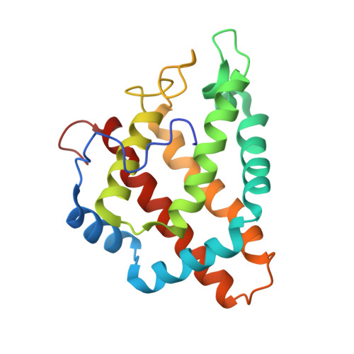
    Protoglobin from Methanosarcina acetivorans C2A (MaPgb), a strictly anaerobic methanogenic Archaea, displays peculiar structural and functional properties within members of the hemoglobin superfamily. In fact, MaPgb-specific loops and a N-terminal extension (20 amino acid residues) completely bury the heme within the protein matrix. Therefore, the access of diatomic gaseous molecules to the heme is granted by two apolar tunnels reaching the heme distal site from locations at the B/G and B/E helix interfaces. The presence of two tunnels within the protein matrix could be partly responsible for the slightly biphasic ligand binding behavior. Unusually, MaPgb oxygenation is favored with respect to carbonylation. Here, the crucial role of Tyr(B10)61 and Ile(G11)149 residues, located in the heme distal site and lining the protein matrix tunnels 1 and 2, respectively, on ligand binding to the heme-Fe-atom and on distal site structural organization is reported. In particular, tunnel 1 accessibility is modulated by a complex reorganization of the Trp(B9)60 and Phe(E11)93 side-chains, triggered by mutations of the Tyr(B10)61 and Ile(G11)149 residues, and affected by the presence and type of the distal heme-bound ligand.


  • Organizational Affiliation

    Department of Physics, University of Genova, Genova, Italy.


Macromolecules
Find similar proteins by:  (by identity cutoff)  |  3D Structure
Entity ID: 1
MoleculeChains Sequence LengthOrganismDetailsImage
Methanosarcina acetivorans protoglobin
A, B
195Methanosarcina acetivoransMutation(s): 2 
Gene Names: MA_2883
UniProt
Find proteins for Q8TLY9 (Methanosarcina acetivorans (strain ATCC 35395 / DSM 2834 / JCM 12185 / C2A))
Explore Q8TLY9 
Go to UniProtKB:  Q8TLY9
Entity Groups  
Sequence Clusters30% Identity50% Identity70% Identity90% Identity95% Identity100% Identity
UniProt GroupQ8TLY9
Sequence Annotations
Expand
  • Reference Sequence
Small Molecules
Ligands 4 Unique
IDChains Name / Formula / InChI Key2D Diagram3D Interactions
HEM
Query on HEM

Download Ideal Coordinates CCD File 
C [auth A],
H [auth B]
PROTOPORPHYRIN IX CONTAINING FE
C34 H32 Fe N4 O4
KABFMIBPWCXCRK-RGGAHWMASA-L
EPE
Query on EPE

Download Ideal Coordinates CCD File 
I [auth B]4-(2-HYDROXYETHYL)-1-PIPERAZINE ETHANESULFONIC ACID
C8 H18 N2 O4 S
JKMHFZQWWAIEOD-UHFFFAOYSA-N
GOL
Query on GOL

Download Ideal Coordinates CCD File 
D [auth A],
E [auth A],
F [auth A]
GLYCEROL
C3 H8 O3
PEDCQBHIVMGVHV-UHFFFAOYSA-N
IPA
Query on IPA

Download Ideal Coordinates CCD File 
G [auth A]ISOPROPYL ALCOHOL
C3 H8 O
KFZMGEQAYNKOFK-UHFFFAOYSA-N
Experimental Data & Validation

Experimental Data

  • Method: X-RAY DIFFRACTION
  • Resolution: 2.20 Å
  • R-Value Free: 0.316 
  • R-Value Work: 0.239 
  • R-Value Observed: 0.243 
  • Space Group: P 1 21 1
Unit Cell:
Length ( Å )Angle ( ˚ )
a = 50.844α = 90
b = 48.368β = 102.5
c = 80.782γ = 90
Software Package:
Software NamePurpose
ADSCdata collection
PHASERphasing
REFMACrefinement
MOSFLMdata reduction
SCALAdata scaling

Structure Validation

View Full Validation Report


Ligand Structure Quality Assessment 


Entry History 

Deposition Data

Revision History  (Full details and data files)

  • Version 1.0: 2011-06-08
    Type: Initial release
  • Version 1.1: 2011-07-13
    Changes: Version format compliance
  • Version 1.2: 2018-01-24
    Changes: Database references
  • Version 1.3: 2023-09-13
    Changes: Data collection, Database references, Derived calculations, Refinement description