


The crystal structure of pyruvate kinase from the hyperthermophilic archaeon Pyrobaculum aerophilum: insights into cooperative regulation
Solomons, J.T.G., Johnson, U., Schoenheit, P., Davies, C.To be published.
Experimental Data Snapshot
wwPDB Validation 3D Report Full Report

Entity ID: 1 | |||||
|---|---|---|---|---|---|
| Molecule | Chains | Sequence Length | Organism | Details | Image |
| Pyruvate kinase | 461 | Pyrobaculum aerophilum str. IM2 | Mutation(s): 0 Gene Names: PAE0819, pyk EC: 2.7.1.40 | 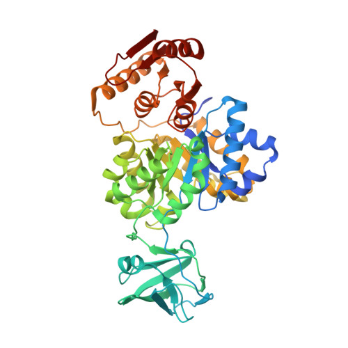 | |
UniProt | |||||
Find proteins for Q8ZYE0 (Pyrobaculum aerophilum (strain ATCC 51768 / DSM 7523 / JCM 9630 / CIP 104966 / NBRC 100827 / IM2)) Explore Q8ZYE0 Go to UniProtKB: Q8ZYE0 | |||||
Entity Groups | |||||
| Sequence Clusters | 30% Identity50% Identity70% Identity90% Identity95% Identity100% Identity | ||||
| UniProt Group | Q8ZYE0 | ||||
Sequence AnnotationsExpand | |||||
| |||||
| Ligands 1 Unique | |||||
|---|---|---|---|---|---|
| ID | Chains | Name / Formula / InChI Key | 2D Diagram | 3D Interactions | |
| SO4 Query on SO4 | C [auth A], D [auth B], E [auth B] | SULFATE ION O4 S QAOWNCQODCNURD-UHFFFAOYSA-L |  | ||
| Length ( Å ) | Angle ( ˚ ) |
|---|---|
| a = 116.3 | α = 90 |
| b = 107.4 | β = 110.5 |
| c = 105 | γ = 90 |
| Software Name | Purpose |
|---|---|
| MAR345 | data collection |
| MOLREP | phasing |
| REFMAC | refinement |
| HKL-2000 | data reduction |
| HKL-2000 | data scaling |
RCSB PDB (citation) is hosted by
RCSB PDB is a member of the

