3QNQ

Crystal structure of the transporter ChbC, the IIC component from the N,N'-diacetylchitobiose-specific phosphotransferase system


Experimental Data Snapshot

  • Method: X-RAY DIFFRACTION
  • Resolution: 3.29 Å
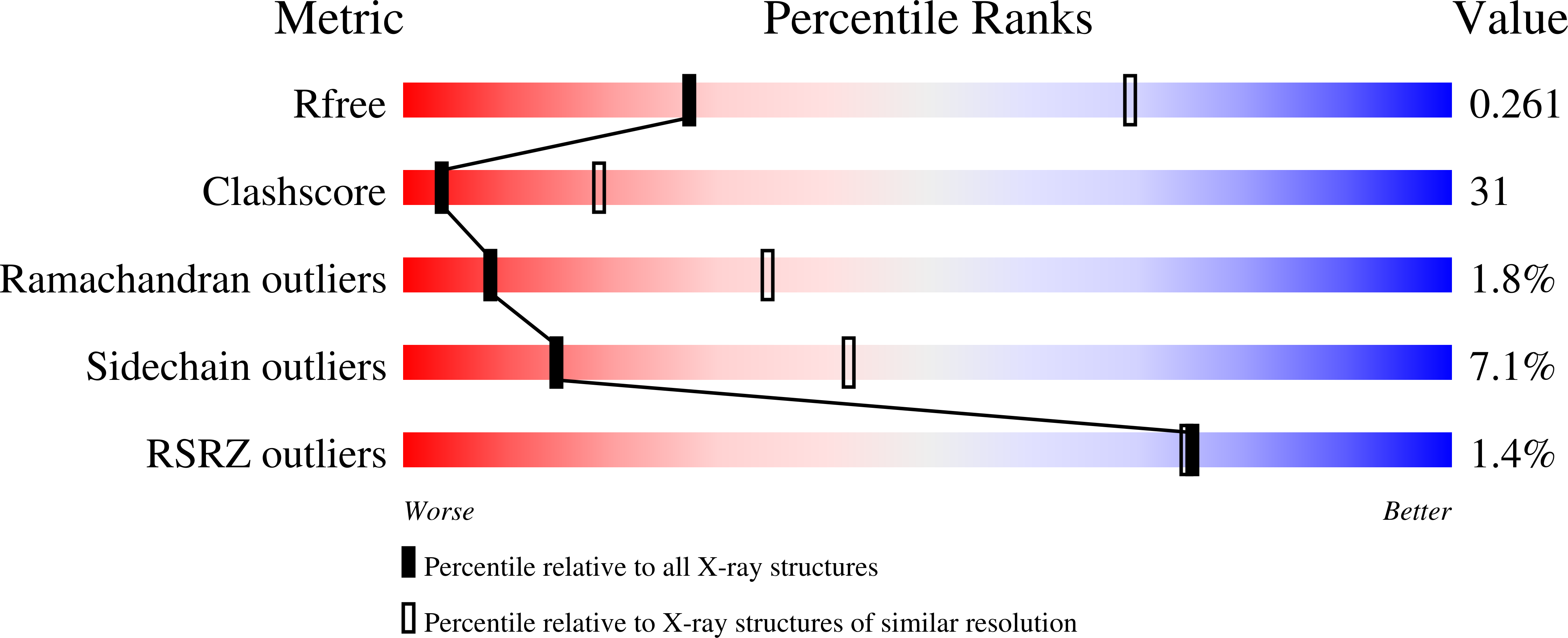
  • R-Value Free: 0.264 
  • R-Value Work: 0.228 
  • R-Value Observed: 0.229 

wwPDB Validation   3D Report Full Report


Ligand Structure Quality Assessment 


This is version 2.1 of the entry. See complete history


Literature

Crystal structure of a phosphorylation-coupled saccharide transporter.

Cao, Y.Jin, X.Levin, E.J.Huang, H.Zong, Y.Quick, M.Weng, J.Pan, Y.Love, J.Punta, M.Rost, B.Hendrickson, W.A.Javitch, J.A.Rajashankar, K.R.Zhou, M.

(2011) Nature 473: 50-54

  • DOI: https://doi.org/10.1038/nature09939
  • Primary Citation of Related Structures:  
    3QNQ

  • PubMed Abstract: 

    Saccharides have a central role in the nutrition of all living organisms. Whereas several saccharide uptake systems are shared between the different phylogenetic kingdoms, the phosphoenolpyruvate-dependent phosphotransferase system exists almost exclusively in bacteria. This multi-component system includes an integral membrane protein EIIC that transports saccharides and assists in their phosphorylation. Here we present the crystal structure of an EIIC from Bacillus cereus that transports diacetylchitobiose. The EIIC is a homodimer, with an expansive interface formed between the amino-terminal halves of the two protomers. The carboxy-terminal half of each protomer has a large binding pocket that contains a diacetylchitobiose, which is occluded from both sides of the membrane with its site of phosphorylation near the conserved His250 and Glu334 residues. The structure shows the architecture of this important class of transporters, identifies the determinants of substrate binding and phosphorylation, and provides a framework for understanding the mechanism of sugar translocation.


  • Organizational Affiliation

    Department of Physiology & Cellular Biophysics, College of Physicians and Surgeons, Columbia University, 630 West 168th Street, New York, New York 10032, USA.


Macromolecules
Find similar proteins by:  (by identity cutoff)  |  3D Structure
Entity ID: 1
MoleculeChains Sequence LengthOrganismDetailsImage
PTS system, cellobiose-specific IIC component
A, B, C, D
442Bacillus cereus ATCC 10987Mutation(s): 0 
Gene Names: BCE_5325celBChbC
EC: 2.7.1.69
Membrane Entity: Yes 
UniProt
Find proteins for Q72XQ0 (Bacillus cereus (strain ATCC 10987 / NRS 248))
Explore Q72XQ0 
Go to UniProtKB:  Q72XQ0
Entity Groups  
Sequence Clusters30% Identity50% Identity70% Identity90% Identity95% Identity100% Identity
UniProt GroupQ72XQ0
Sequence Annotations
Expand
  • Reference Sequence
Oligosaccharides

Help

Entity ID: 2
MoleculeChains Length2D Diagram Glycosylation3D Interactions
2-acetamido-2-deoxy-beta-D-glucopyranose-(1-4)-2-acetamido-2-deoxy-beta-D-glucopyranose
E, G, I, K
2N/A
Glycosylation Resources
GlyTouCan:  G42666HT
GlyCosmos:  G42666HT
GlyGen:  G42666HT
Entity ID: 3
MoleculeChains Length2D Diagram Glycosylation3D Interactions
alpha-D-glucopyranose-(1-4)-alpha-D-glucopyranose
F, H, J, L
2N/A
Glycosylation Resources
GlyTouCan:  G07411ON
GlyCosmos:  G07411ON
Small Molecules
Ligands 2 Unique
IDChains Name / Formula / InChI Key2D Diagram3D Interactions
ZDM
Query on ZDM

Download Ideal Coordinates CCD File 
M [auth A],
O [auth A],
R [auth C],
U [auth C]
nonyl 4-O-alpha-D-glucopyranosyl-beta-D-glucopyranoside
C21 H40 O11
KCCBGPCYGBPHBR-ZESVGKPKSA-N
CIT
Query on CIT

Download Ideal Coordinates CCD File 
N [auth A]
P [auth B]
Q [auth B]
S [auth C]
T [auth C]
N [auth A],
P [auth B],
Q [auth B],
S [auth C],
T [auth C],
V [auth C],
W [auth D]
CITRIC ACID
C6 H8 O7
KRKNYBCHXYNGOX-UHFFFAOYSA-N
Experimental Data & Validation

Experimental Data

  • Method: X-RAY DIFFRACTION
  • Resolution: 3.29 Å
  • R-Value Free: 0.264 
  • R-Value Work: 0.228 
  • R-Value Observed: 0.229 
  • Space Group: P 43 21 2
Unit Cell:
Length ( Å )Angle ( ˚ )
a = 132.812α = 90
b = 132.812β = 90
c = 452.954γ = 90
Software Package:
Software NamePurpose
DENZOdata reduction
SCALEPACKdata scaling
SHELXphasing
DMphasing
PHENIXrefinement
PDB_EXTRACTdata extraction
HKL-2000data collection
HKL-2000data reduction
HKL-2000data scaling
SHARPphasing

Structure Validation

View Full Validation Report


Ligand Structure Quality Assessment 


Entry History 

Deposition Data

Revision History  (Full details and data files)

  • Version 1.0: 2011-04-06
    Type: Initial release
  • Version 1.1: 2011-07-13
    Changes: Version format compliance
  • Version 1.2: 2017-11-08
    Changes: Advisory, Refinement description
  • Version 2.0: 2020-07-29
    Type: Remediation
    Reason: Carbohydrate remediation
    Changes: Advisory, Atomic model, Data collection, Database references, Derived calculations, Non-polymer description, Structure summary
  • Version 2.1: 2024-02-21
    Changes: Data collection, Database references, Structure summary