


Structure of Proteasome Tether
Schumacher, M.A., Glover, T., Weijun, X.To be published.
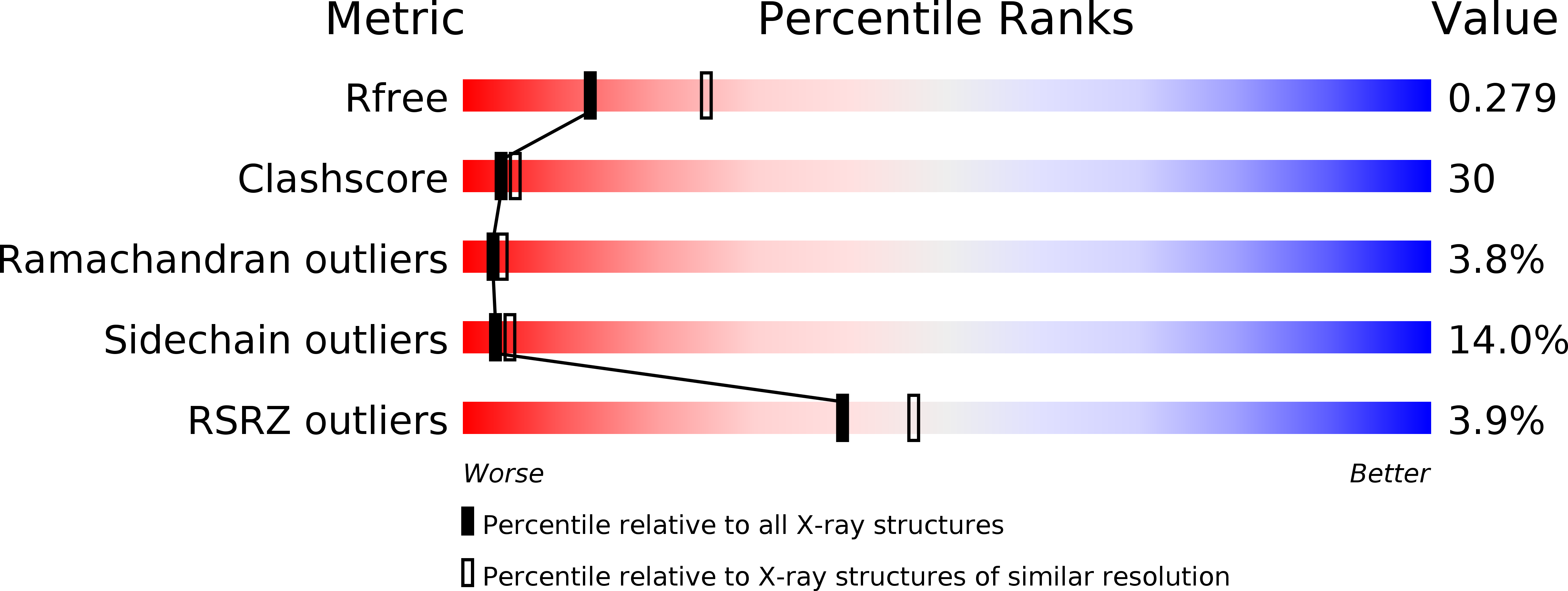
Experimental Data Snapshot
wwPDB Validation 3D Report Full Report
Entity ID: 1 | |||||
|---|---|---|---|---|---|
| Molecule | Chains | Sequence Length | Organism | Details | Image |
| Protein cut8 | 245 | Schizosaccharomyces pombe | Mutation(s): 0 Gene Names: cut8, SPAC17C9.13c | 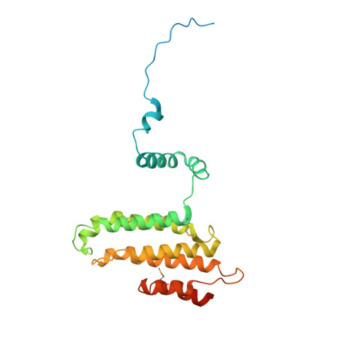 | |
UniProt | |||||
Find proteins for P38937 (Schizosaccharomyces pombe (strain 972 / ATCC 24843)) Explore P38937 Go to UniProtKB: P38937 | |||||
Entity Groups | |||||
| Sequence Clusters | 30% Identity50% Identity70% Identity90% Identity95% Identity100% Identity | ||||
| UniProt Group | P38937 | ||||
Sequence AnnotationsExpand | |||||
| |||||
| Modified Residues 1 Unique | |||||
|---|---|---|---|---|---|
| ID | Chains | Type | Formula | 2D Diagram | Parent |
| MSE Query on MSE | A, B | L-PEPTIDE LINKING | C5 H11 N O2 Se |  | MET |
| Length ( Å ) | Angle ( ˚ ) |
|---|---|
| a = 35.66 | α = 103.92 |
| b = 51.18 | β = 100.02 |
| c = 71.33 | γ = 98 |
| Software Name | Purpose |
|---|---|
| ADSC | data collection |
| SOLVE | phasing |
| CNS | refinement |
| MOSFLM | data reduction |
| SCALA | data scaling |
RCSB PDB (citation) is hosted by
RCSB PDB is a member of the

