


Crystal structure of human PACSIN 2 F-BAR domain
Bai, X., Meng, G., Zheng, X.To be published.
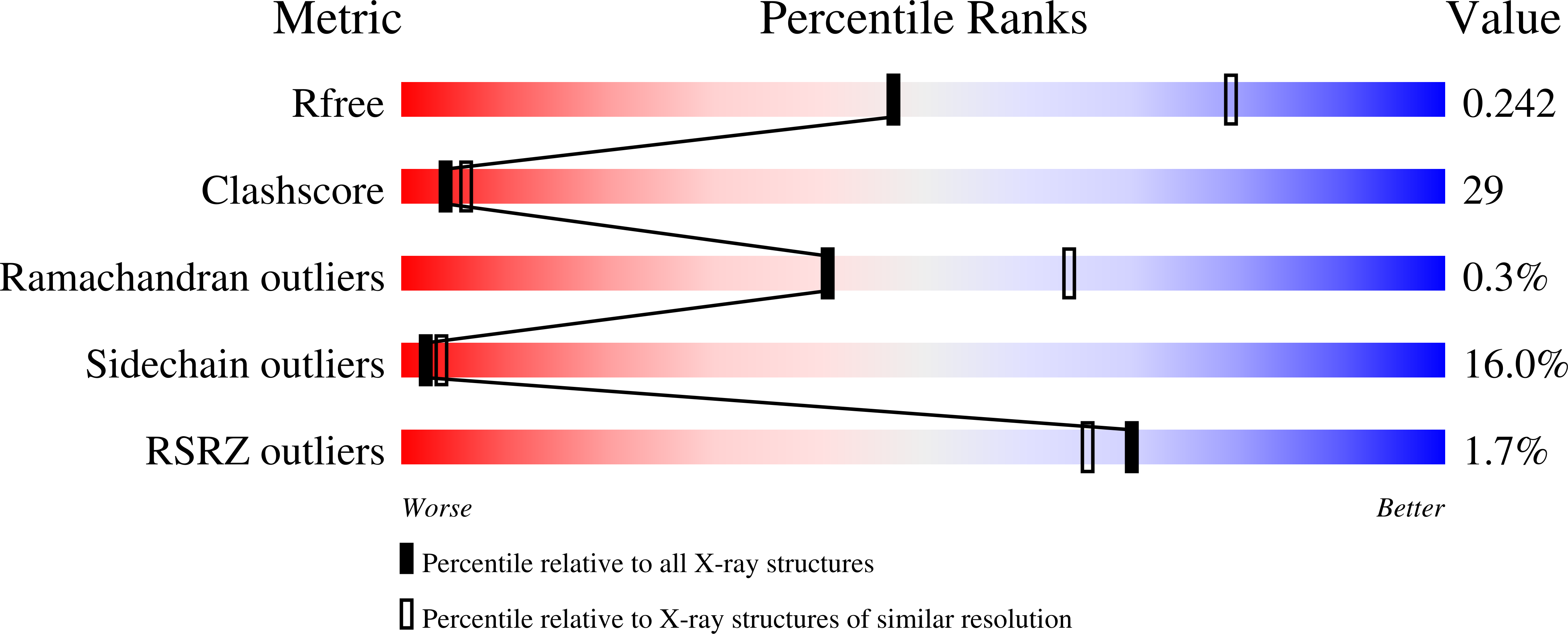
Experimental Data Snapshot
wwPDB Validation 3D Report Full Report
Entity ID: 1 | |||||
|---|---|---|---|---|---|
| Molecule | Chains | Sequence Length | Organism | Details | Image |
| Protein kinase C and casein kinase substrate in neurons protein 2 | 289 | Homo sapiens | Mutation(s): 0 Gene Names: PACSIN2 Membrane Entity: Yes |  | |
UniProt & NIH Common Fund Data Resources | |||||
Find proteins for Q9UNF0 (Homo sapiens) Explore Q9UNF0 Go to UniProtKB: Q9UNF0 | |||||
PHAROS: Q9UNF0 GTEx: ENSG00000100266 | |||||
Entity Groups | |||||
| Sequence Clusters | 30% Identity50% Identity70% Identity90% Identity95% Identity100% Identity | ||||
| UniProt Group | Q9UNF0 | ||||
Sequence AnnotationsExpand | |||||
| |||||
| Ligands 1 Unique | |||||
|---|---|---|---|---|---|
| ID | Chains | Name / Formula / InChI Key | 2D Diagram | 3D Interactions | |
| CA Query on CA | E [auth A], F [auth B], G [auth C], H [auth D] | CALCIUM ION Ca BHPQYMZQTOCNFJ-UHFFFAOYSA-N |  | ||
| Length ( Å ) | Angle ( ˚ ) |
|---|---|
| a = 31.562 | α = 90 |
| b = 353.588 | β = 90.02 |
| c = 86.045 | γ = 90 |
| Software Name | Purpose |
|---|---|
| HKL-2000 | data collection |
| AMoRE | phasing |
| REFMAC | refinement |
| MOSFLM | data reduction |
| SCALA | data scaling |
RCSB PDB (citation) is hosted by
RCSB PDB is a member of the

