3PKY

Polymerase Domain from Mycobacterium tuberculosis Ligase D in complex with DNA, UTP and Manganese.


Experimental Data Snapshot

  • Method: X-RAY DIFFRACTION
  • Resolution: 3.10 Å
  • R-Value Free: 0.248 
  • R-Value Work: 0.191 
  • R-Value Observed: 0.194 

wwPDB Validation   3D Report Full Report


Ligand Structure Quality Assessment 


This is version 1.2 of the entry. See complete history


Literature

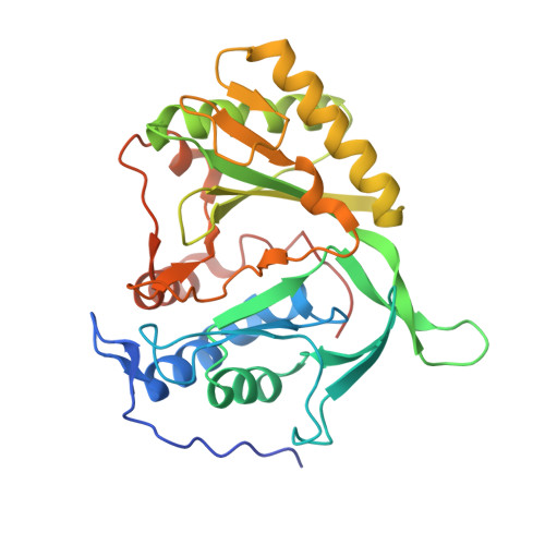
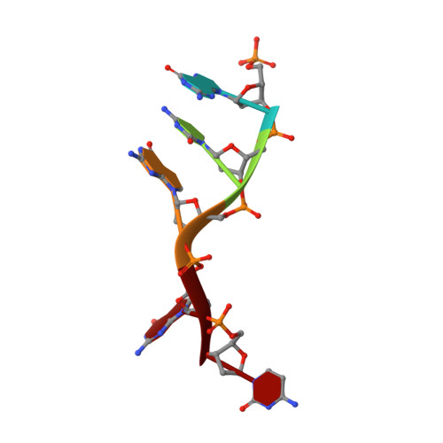
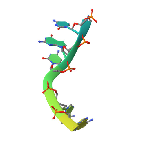
Structure of a Preternary Complex Involving a Prokaryotic NHEJ DNA Polymerase.

Brissett, N.C.Martin, M.J.Pitcher, R.S.Bianchi, J.Juarez, R.Green, A.J.Fox, G.C.Blanco, L.Doherty, A.J.

(2011) Mol Cell 41: 221-231

  • DOI: https://doi.org/10.1016/j.molcel.2010.12.026
  • Primary Citation of Related Structures:  
    3PKY

  • PubMed Abstract: 

    In many prokaryotes, a specific DNA primase/polymerase (PolDom) is required for nonhomologous end joining (NHEJ) repair of DNA double-strand breaks (DSBs). Here, we report the crystal structure of a catalytically active conformation of Mycobacterium tuberculosis PolDom, consisting of a polymerase bound to a DNA end with a 3' overhang, two metal ions, and an incoming nucleotide but, significantly, lacking a primer strand. This structure represents a polymerase:DNA complex in a preternary intermediate state. This polymerase complex occurs in solution, stabilizing the enzyme on DNA ends and promoting nucleotide extension of short incoming termini. We also demonstrate that the invariant Arg(220), contained in a conserved loop (loop 2), plays an essential role in catalysis by regulating binding of a second metal ion in the active site. We propose that this NHEJ intermediate facilitates extension reactions involving critically short or noncomplementary DNA ends, thus promoting break repair and minimizing sequence loss during DSB repair.


  • Organizational Affiliation

    Genome Damage and Stability Centre, University of Sussex, Brighton BN1 9RQ, UK.


Macromolecules

Find similar proteins by:  (by identity cutoff)  |  3D Structure
Entity ID: 1
MoleculeChains Sequence LengthOrganismDetailsImage
Putative DNA ligase-like protein
A, B
303Mycobacterium tuberculosisMutation(s): 0 
Gene Names: MT0965MTCY08D9.01cMTCY10D7.36cRv0938
EC: 2.7.7
UniProt
Find proteins for P9WNV3 (Mycobacterium tuberculosis (strain ATCC 25618 / H37Rv))
Explore P9WNV3 
Go to UniProtKB:  P9WNV3
Entity Groups  
Sequence Clusters30% Identity50% Identity70% Identity90% Identity95% Identity100% Identity
UniProt GroupP9WNV3
Sequence Annotations
Expand
  • Reference Sequence

Find similar nucleic acids by:  Sequence   |   3D Structure  

Entity ID: 2
MoleculeChains LengthOrganismImage
DNA 5'-D(P*GP*CP*GP*GP*C)-3'5N/A
Sequence Annotations
Expand
  • Reference Sequence

Find similar nucleic acids by:  Sequence   |   3D Structure  

Entity ID: 3
MoleculeChains LengthOrganismImage
DNA 5'-D(*G*CP*CP*GP*CP*AP*AP*CP*GP*CP*AP*CP*G)-3'13N/A
Sequence Annotations
Expand
  • Reference Sequence
Experimental Data & Validation

Experimental Data

  • Method: X-RAY DIFFRACTION
  • Resolution: 3.10 Å
  • R-Value Free: 0.248 
  • R-Value Work: 0.191 
  • R-Value Observed: 0.194 
  • Space Group: P 41
Unit Cell:
Length ( Å )Angle ( ˚ )
a = 145.711α = 90
b = 145.711β = 90
c = 44.96γ = 90
Software Package:
Software NamePurpose
DENZOdata reduction
SCALEPACKdata scaling
PHASERphasing
REFMACrefinement
PDB_EXTRACTdata extraction

Structure Validation

View Full Validation Report


Ligand Structure Quality Assessment 


Entry History 

Deposition Data

Revision History  (Full details and data files)

  • Version 1.0: 2011-02-16
    Type: Initial release
  • Version 1.1: 2011-07-13
    Changes: Version format compliance
  • Version 1.2: 2024-02-21
    Changes: Data collection, Database references, Derived calculations