3PKW

Crystal Structure of Toxoflavin Lyase (TflA) bound to Mn(II)


Experimental Data Snapshot

  • Method: X-RAY DIFFRACTION
  • Resolution: 1.80 Å
  • R-Value Free: 0.223 
  • R-Value Work: 0.189 
  • R-Value Observed: 0.191 

wwPDB Validation   3D Report Full Report


This is version 1.4 of the entry. See complete history


Literature

Toxoflavin lyase requires a novel 1-his-2-carboxylate facial triad .

Fenwick, M.K.Philmus, B.Begley, T.P.Ealick, S.E.

(2011) Biochemistry 50: 1091-1100

  • DOI: https://doi.org/10.1021/bi101741v
  • Primary Citation of Related Structures:  
    3PKV, 3PKW, 3PKX

  • PubMed Abstract: 

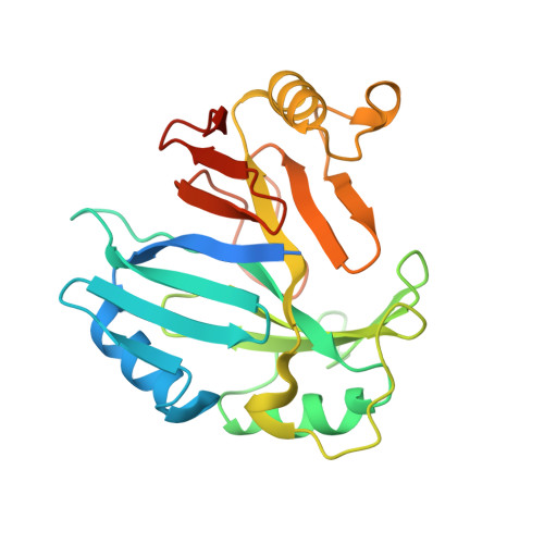
    High-resolution crystal structures are reported for apo, holo, and substrate-bound forms of a toxoflavin-degrading metalloenzyme (TflA). In addition, the degradation reaction is shown to be dependent on oxygen, Mn(II), and dithiothreitol in vitro. Despite its low sequence identity with proteins of known structure, TflA is structurally homologous to proteins of the vicinal oxygen chelate superfamily. Like other metalloenzymes in this superfamily, the TflA fold contains four modules that associate to form a metal binding site; however, the fold displays a rare rearrangement of the structural modules indicative of domain permutation. Moreover, unlike the 2-His-1-carboxylate facial triad commonly utilized by vicinal oxygen chelate dioxygenases and other dioxygen-activating non-heme Fe(II) enzymes, the metal center in TflA consists of a 1-His-2-carboxylate facial triad. The substrate-bound complex shows square-pyramidal geometry in which one position is occupied by O5 of toxoflavin. The open coordination site is predicted to be the dioxygen binding site. TflA appears to stabilize the reduced form of toxoflavin through second-sphere interactions. This anionic species is predicted to be the electron source responsible for reductive activation of oxygen to produce a peroxytoxoflavin intermediate.


  • Organizational Affiliation

    Department of Chemistry and Chemical Biology, Cornell University, Ithaca, New York 14853, United States.


Macromolecules
Find similar proteins by:  (by identity cutoff)  |  3D Structure
Entity ID: 1
MoleculeChains Sequence LengthOrganismDetailsImage
Toxoflavin lyase (TflA)252Paenibacillus polymyxaMutation(s): 0 
Gene Names: tflA
UniProt
Find proteins for E3SET7 (Paenibacillus polymyxa)
Explore E3SET7 
Go to UniProtKB:  E3SET7
Entity Groups  
Sequence Clusters30% Identity50% Identity70% Identity90% Identity95% Identity100% Identity
UniProt GroupE3SET7
Sequence Annotations
Expand
  • Reference Sequence
Small Molecules
Ligands 1 Unique
IDChains Name / Formula / InChI Key2D Diagram3D Interactions
MN
Query on MN

Download Ideal Coordinates CCD File 
B [auth A]MANGANESE (II) ION
Mn
WAEMQWOKJMHJLA-UHFFFAOYSA-N
Experimental Data & Validation

Experimental Data

  • Method: X-RAY DIFFRACTION
  • Resolution: 1.80 Å
  • R-Value Free: 0.223 
  • R-Value Work: 0.189 
  • R-Value Observed: 0.191 
  • Space Group: H 3
Unit Cell:
Length ( Å )Angle ( ˚ )
a = 110.856α = 90
b = 110.856β = 90
c = 55.734γ = 120
Software Package:
Software NamePurpose
DENZOdata reduction
SCALEPACKdata scaling
SHELXphasing
PHENIXrefinement
PDB_EXTRACTdata extraction
ADSCdata collection
HKL-2000data reduction
HKL-2000data scaling
SHELXDphasing

Structure Validation

View Full Validation Report


Entry History 

Deposition Data

Revision History  (Full details and data files)

  • Version 1.0: 2011-01-12
    Type: Initial release
  • Version 1.1: 2011-07-13
    Changes: Version format compliance
  • Version 1.2: 2011-11-16
    Changes: Atomic model
  • Version 1.3: 2017-11-08
    Changes: Refinement description
  • Version 1.4: 2024-02-21
    Changes: Data collection, Database references, Derived calculations