3PEL

Structure of Greyhound Hemoglobin: Origin of High Oxygen Affinity


Experimental Data Snapshot

  • Method: X-RAY DIFFRACTION
  • Resolution: 1.90 Å
  • R-Value Free: 0.232 
  • R-Value Work: 0.188 
  • R-Value Observed: 0.190 

wwPDB Validation   3D Report Full Report


Ligand Structure Quality Assessment 


This is version 1.2 of the entry. See complete history


Literature

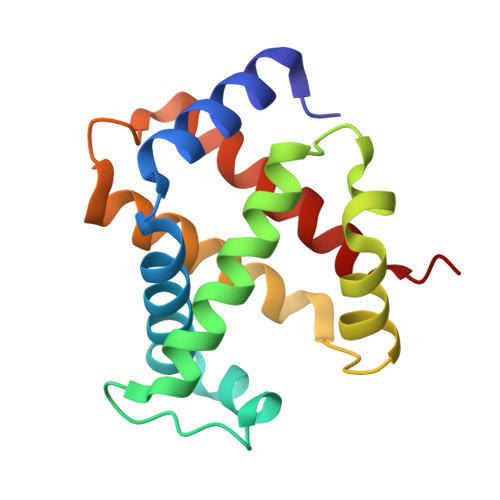
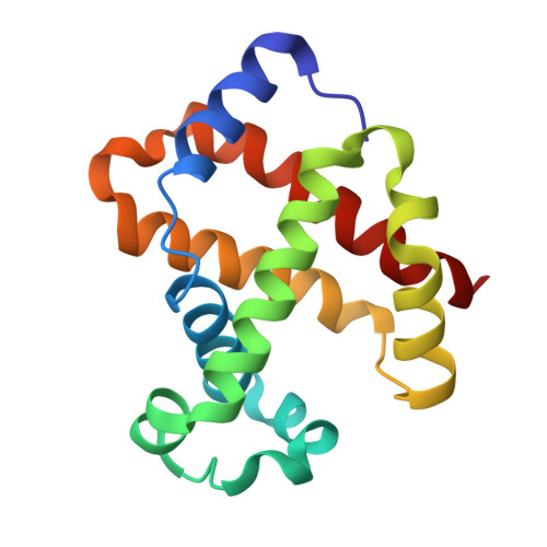
Structure of Greyhound hemoglobin: origin of high oxygen affinity.

Bhatt, V.S.Zaldivar-Lopez, S.Harris, D.R.Couto, C.G.Wang, P.G.Palmer, A.F.

(2011) Acta Crystallogr D Biol Crystallogr 67: 395-402

  • DOI: https://doi.org/10.1107/S0907444911006044
  • Primary Citation of Related Structures:  
    3PEL

  • PubMed Abstract: 

    This study presents the crystal structure of Greyhound hemoglobin (GrHb) determined to 1.9 Å resolution. GrHb was found to crystallize with an α₁β₁ dimer in the asymmetric unit and belongs to the R2 state. Oxygen-affinity measurements combined with the fact that GrHb crystallizes in the R2 state despite the high-salt conditions used for crystallization strongly indicate that GrHb can serve as a model high-oxygen-affinity hemoglobin (Hb) for higher mammals, especially humans. Structural analysis of GrHb and its comparison with the R2-state of human Hb revealed several regions that can potentially contribute to the high oxygen affinity of GrHb and serve to rationalize the additional stability of the R2-state of GrHb. A previously well studied hydrophobic cluster of bar-headed goose Hb near α119 was also incorporated in the comparison between GrHb and human Hb. Finally, a structural comparison with generic dog Hb and maned wolf Hb was conducted, revealing that in contrast to GrHb these structures belong to the R state of Hb and raising the intriguing possibility of an additional allosteric factor co-purifying with GrHb that can modulate its quaternary structure.


  • Organizational Affiliation

    Biophysics Graduate Program, The Ohio State University, Columbus, OH 43210, USA.


Macromolecules
Find similar proteins by:  (by identity cutoff)  |  3D Structure
Entity ID: 1
MoleculeChains Sequence LengthOrganismDetailsImage
Hemoglobin subunit alpha141Canis lupus familiarisMutation(s): 0 
UniProt
Find proteins for P60529 (Canis lupus familiaris)
Explore P60529 
Go to UniProtKB:  P60529
Entity Groups  
Sequence Clusters30% Identity50% Identity70% Identity90% Identity95% Identity100% Identity
UniProt GroupP60529
Sequence Annotations
Expand
  • Reference Sequence
Find similar proteins by:  (by identity cutoff)  |  3D Structure
Entity ID: 2
MoleculeChains Sequence LengthOrganismDetailsImage
Hemoglobin subunit beta146Canis lupus familiarisMutation(s): 0 
UniProt
Find proteins for P60524 (Canis lupus familiaris)
Explore P60524 
Go to UniProtKB:  P60524
Entity Groups  
Sequence Clusters30% Identity50% Identity70% Identity90% Identity95% Identity100% Identity
UniProt GroupP60524
Sequence Annotations
Expand
  • Reference Sequence
Small Molecules
Ligands 1 Unique
IDChains Name / Formula / InChI Key2D Diagram3D Interactions
HEM
Query on HEM

Download Ideal Coordinates CCD File 
C [auth A],
D [auth B]
PROTOPORPHYRIN IX CONTAINING FE
C34 H32 Fe N4 O4
KABFMIBPWCXCRK-RGGAHWMASA-L
Experimental Data & Validation

Experimental Data

  • Method: X-RAY DIFFRACTION
  • Resolution: 1.90 Å
  • R-Value Free: 0.232 
  • R-Value Work: 0.188 
  • R-Value Observed: 0.190 
  • Space Group: C 1 2 1
Unit Cell:
Length ( Å )Angle ( ˚ )
a = 87.973α = 90
b = 88.045β = 103.37
c = 53.073γ = 90
Software Package:
Software NamePurpose
CrystalCleardata collection
AMoREphasing
REFMACrefinement
CrystalCleardata reduction
CrystalCleardata scaling

Structure Validation

View Full Validation Report


Ligand Structure Quality Assessment 


Entry History 

Deposition Data

Revision History  (Full details and data files)

  • Version 1.0: 2010-11-03
    Type: Initial release
  • Version 1.1: 2011-07-13
    Changes: Version format compliance
  • Version 1.2: 2023-09-06
    Changes: Data collection, Database references, Derived calculations, Refinement description