3PAC

Crystal structure of PduT a trimeric bacterial microcompartment protein with 4Fe-4S cluster binding site


Experimental Data Snapshot

  • Method: X-RAY DIFFRACTION
  • Resolution: 1.86 Å
  • R-Value Free: 0.252 
  • R-Value Work: 0.199 
  • R-Value Observed: 0.204 

wwPDB Validation   3D Report Full Report


This is version 1.2 of the entry. See complete history


Literature

Structure of PduT, a trimeric bacterial microcompartment protein with a 4Fe-4S cluster-binding site

Pang, A.Warren, M.J.Pickersgill, R.W.

(2011) Acta Crystallogr D Biol Crystallogr 67: 91-96

  • DOI: https://doi.org/10.1107/S0907444910050201
  • Primary Citation of Related Structures:  
    3PAC

  • PubMed Abstract: 

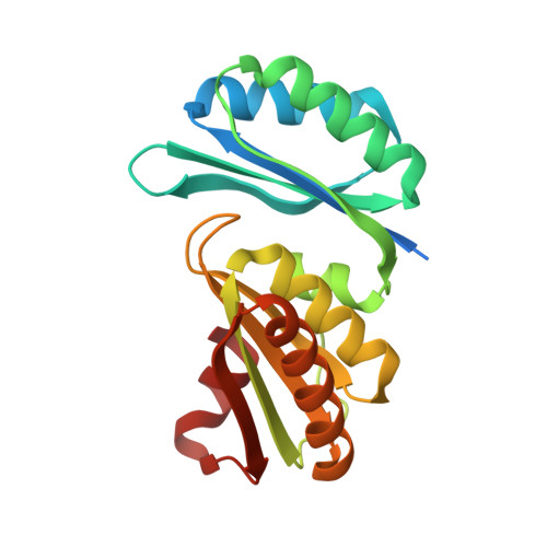
    Propanediol metabolism in Citrobacter freundii occurs within a metabolosome, a subcellular proteinaceous bacterial microcompartment. The propanediol-utilization (Pdu) microcompartment shell is constructed from thousands of hexagonal-shaped protein complexes made from seven different types of protein subunit. Here, the structure of the bacterial microcompartment protein PduT, which has a tandem structural repeat within the subunit and forms trimers with pseudo-hexagonal symmetry, is reported. This trimeric assembly forms a flat approximately hexagonally shaped disc with a central pore that is suitable for a 4Fe-4S cluster. The essentially cubic shaped 4Fe-4S cluster conforms to the threefold symmetry of the trimer with one free iron, the role of which could be to supply electrons to an associated microcompartment enzyme, PduS.


  • Organizational Affiliation

    School of Biological and Chemical Sciences, Queen Mary University of London, Mile End Road, London E1 4NS, England.


Macromolecules
Find similar proteins by:  (by identity cutoff)  |  3D Structure
Entity ID: 1
MoleculeChains Sequence LengthOrganismDetailsImage
Shell protein204Citrobacter freundiiMutation(s): 0 
Gene Names: pduT
UniProt
Find proteins for B1VB78 (Citrobacter freundii)
Explore B1VB78 
Go to UniProtKB:  B1VB78
Entity Groups  
Sequence Clusters30% Identity50% Identity70% Identity90% Identity95% Identity100% Identity
UniProt GroupB1VB78
Sequence Annotations
Expand
  • Reference Sequence
Experimental Data & Validation

Experimental Data

  • Method: X-RAY DIFFRACTION
  • Resolution: 1.86 Å
  • R-Value Free: 0.252 
  • R-Value Work: 0.199 
  • R-Value Observed: 0.204 
  • Space Group: P 63
Unit Cell:
Length ( Å )Angle ( ˚ )
a = 67.68α = 90
b = 67.68β = 90
c = 62.03γ = 120
Software Package:
Software NamePurpose
ADSCdata collection
PHENIXmodel building
REFMACrefinement
MOSFLMdata reduction
SCALAdata scaling
PHENIXphasing

Structure Validation

View Full Validation Report


Entry History 

Deposition Data

Revision History  (Full details and data files)

  • Version 1.0: 2010-12-01
    Type: Initial release
  • Version 1.1: 2011-07-13
    Changes: Version format compliance
  • Version 1.2: 2024-03-20
    Changes: Data collection, Database references