3OYX

Haloferax volcanii Malate Synthase Magnesium/Glyoxylate Complex


Experimental Data Snapshot

  • Method: X-RAY DIFFRACTION
  • Resolution: 2.51 Å
  • R-Value Free: 0.248 
  • R-Value Work: 0.192 
  • R-Value Observed: 0.195 

wwPDB Validation   3D Report Full Report


This is version 1.3 of the entry. See complete history


Literature

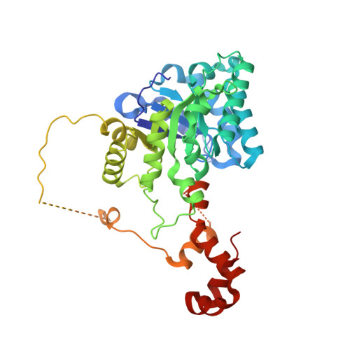
Crystal structures of a halophilic archaeal malate synthase from Haloferax volcanii and comparisons with isoforms A and G.

Bracken, C.D.Neighbor, A.M.Lamlenn, K.K.Thomas, G.C.Schubert, H.L.Whitby, F.G.Howard, B.R.

(2011) BMC Struct Biol 11: 23-23

  • DOI: https://doi.org/10.1186/1472-6807-11-23
  • Primary Citation of Related Structures:  
    3OYX, 3OYZ, 3PUG

  • PubMed Abstract: 

    Malate synthase, one of the two enzymes unique to the glyoxylate cycle, is found in all three domains of life, and is crucial to the utilization of two-carbon compounds for net biosynthetic pathways such as gluconeogenesis. In addition to the main isoforms A and G, so named because of their differential expression in E. coli grown on either acetate or glycolate respectively, a third distinct isoform has been identified. These three isoforms differ considerably in size and sequence conservation. The A isoform (MSA) comprises ~530 residues, the G isoform (MSG) is ~730 residues, and this third isoform (MSH-halophilic) is ~430 residues in length. Both isoforms A and G have been structurally characterized in detail, but no structures have been reported for the H isoform which has been found thus far only in members of the halophilic Archaea.


  • Organizational Affiliation

    Department of Physical Science, Southern Utah University, Cedar City, UT 84720-2470, USA.


Macromolecules
Find similar proteins by:  (by identity cutoff)  |  3D Structure
Entity ID: 1
MoleculeChains Sequence LengthOrganismDetailsImage
Malate synthase433Haloferax volcanii DS2Mutation(s): 0 
EC: 2.3.3.9
UniProt
Find proteins for D4GTL2 (Haloferax volcanii (strain ATCC 29605 / DSM 3757 / JCM 8879 / NBRC 14742 / NCIMB 2012 / VKM B-1768 / DS2))
Explore D4GTL2 
Go to UniProtKB:  D4GTL2
Entity Groups  
Sequence Clusters30% Identity50% Identity70% Identity90% Identity95% Identity100% Identity
UniProt GroupD4GTL2
Sequence Annotations
Expand
  • Reference Sequence
Small Molecules
Ligands 4 Unique
IDChains Name / Formula / InChI Key2D Diagram3D Interactions
GLV
Query on GLV

Download Ideal Coordinates CCD File 
B [auth A],
C [auth A]
GLYOXYLIC ACID
C2 H2 O3
HHLFWLYXYJOTON-UHFFFAOYSA-N
K
Query on K

Download Ideal Coordinates CCD File 
J [auth A],
K [auth A],
L [auth A]
POTASSIUM ION
K
NPYPAHLBTDXSSS-UHFFFAOYSA-N
CL
Query on CL

Download Ideal Coordinates CCD File 
E [auth A],
F [auth A],
G [auth A],
H [auth A],
I [auth A]
CHLORIDE ION
Cl
VEXZGXHMUGYJMC-UHFFFAOYSA-M
MG
Query on MG

Download Ideal Coordinates CCD File 
D [auth A]MAGNESIUM ION
Mg
JLVVSXFLKOJNIY-UHFFFAOYSA-N
Experimental Data & Validation

Experimental Data

  • Method: X-RAY DIFFRACTION
  • Resolution: 2.51 Å
  • R-Value Free: 0.248 
  • R-Value Work: 0.192 
  • R-Value Observed: 0.195 
  • Space Group: H 3 2
Unit Cell:
Length ( Å )Angle ( ˚ )
a = 155.023α = 90
b = 155.023β = 90
c = 141.787γ = 120
Software Package:
Software NamePurpose
DENZOdata reduction
SCALEPACKdata scaling
PHASERphasing
REFMACrefinement
PDB_EXTRACTdata extraction

Structure Validation

View Full Validation Report


Entry History 

Deposition Data

Revision History  (Full details and data files)

  • Version 1.0: 2011-06-01
    Type: Initial release
  • Version 1.1: 2011-07-13
    Changes: Version format compliance
  • Version 1.2: 2017-11-08
    Changes: Refinement description
  • Version 1.3: 2024-02-21
    Changes: Data collection, Database references, Derived calculations, Structure summary