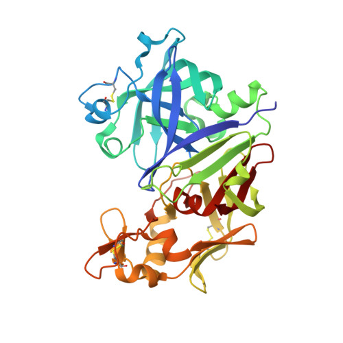
3OQF

Crystal Structure Analysis of Renin-indole-piperazine inhibitor complexes


Experimental Data Snapshot

  • Method: X-RAY DIFFRACTION
  • Resolution: 2.78 Å
  • R-Value Free: 0.201 
  • R-Value Work: 0.180 
  • R-Value Observed: 0.181 

wwPDB Validation   3D Report Full Report


Ligand Structure Quality Assessment 


This is version 1.2 of the entry. See complete history


Literature

Discovery and optimization of a new class of potent and non-chiral indole-3-carboxamide-based renin inhibitors.

Scheiper, B.Matter, H.Steinhagen, H.Stilz, U.Bocskei, Z.Fleury, V.McCort, G.

(2010) Bioorg Med Chem Lett 20: 6268-6272

  • DOI: https://doi.org/10.1016/j.bmcl.2010.08.092
  • Primary Citation of Related Structures:  
    3OOT, 3OQF, 3OQK

  • PubMed Abstract: 

    Selective inhibition of the aspartyl protease renin has gained attraction as an interesting approach to control hypertension and associated cardiovascular risk factors given its unique position in the renin-angiotensin system. Using a combination of high-throughput screening, parallel synthesis, X-ray crystallography and structure-based design, we identified and optimized a novel series of potent and non-chiral indole-3-carboxamides with remarkable potency for renin. The most potent compound 5k displays an IC(50) value of 2nM.


  • Organizational Affiliation

    Sanofi-Aventis Deutschland GmbH, Chemical and Analytical Sciences, Building G878, D-65926 Frankfurt, Germany. Bodo.Scheiper@sanofi-aventis.com


Macromolecules
Find similar proteins by:  (by identity cutoff)  |  3D Structure
Entity ID: 1
MoleculeChains Sequence LengthOrganismDetailsImage
Renin
A, B
340Homo sapiensMutation(s): 0 
Gene Names: REN
EC: 3.4.23.15
UniProt & NIH Common Fund Data Resources
Find proteins for P00797 (Homo sapiens)
Explore P00797 
Go to UniProtKB:  P00797
PHAROS:  P00797
GTEx:  ENSG00000143839 
Entity Groups  
Sequence Clusters30% Identity50% Identity70% Identity90% Identity95% Identity100% Identity
UniProt GroupP00797
Sequence Annotations
Expand
  • Reference Sequence
Small Molecules
Binding Affinity Annotations 
IDSourceBinding Affinity
S51 BindingDB:  3OQF IC50: 91 (nM) from 1 assay(s)
PDBBind:  3OQF IC50: 91 (nM) from 1 assay(s)
Experimental Data & Validation

Experimental Data

  • Method: X-RAY DIFFRACTION
  • Resolution: 2.78 Å
  • R-Value Free: 0.201 
  • R-Value Work: 0.180 
  • R-Value Observed: 0.181 
  • Space Group: P 21 3
Unit Cell:
Length ( Å )Angle ( ˚ )
a = 143.22α = 90
b = 143.22β = 90
c = 143.22γ = 90
Software Package:
Software NamePurpose
SCALEPACKdata scaling
BUSTER-TNTrefinement
PDB_EXTRACTdata extraction
CrystalCleardata collection
MOSFLMdata reduction
BUSTERrefinement

Structure Validation

View Full Validation Report


Ligand Structure Quality Assessment 


Entry History 

Deposition Data

  • Released Date: 2010-10-13 
  • Deposition Author(s): Bocskei, Z.

Revision History  (Full details and data files)

  • Version 1.0: 2010-10-13
    Type: Initial release
  • Version 1.1: 2011-07-13
    Changes: Version format compliance
  • Version 1.2: 2020-07-29
    Type: Remediation
    Reason: Carbohydrate remediation
    Changes: Data collection, Derived calculations, Structure summary