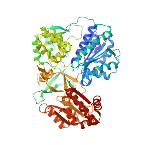
Conformational Changes of NADPH-Cytochrome P450 Oxidoreductase Are Essential for Catalysis and Cofactor Binding.
Xia, C., Hamdane, D., Shen, A.L., Choi, V., Kasper, C.B., Pearl, N.M., Zhang, H., Im, S.C., Waskell, L., Kim, J.J.(2011) J Biol Chem 286: 16246-16260
- PubMed: 21345800
- DOI: https://doi.org/10.1074/jbc.M111.230532
- Primary Citation of Related Structures:
3OJW, 3OJX - PubMed Abstract:
The crystal structure of NADPH-cytochrome P450 reductase (CYPOR) implies that a large domain movement is essential for electron transfer from NADPH via FAD and FMN to its redox partners. To test this hypothesis, a disulfide bond was engineered between residues Asp(147) and Arg(514) in the FMN and FAD domains, respectively. The cross-linked form of this mutant protein, designated 147CC514, exhibited a significant decrease in the rate of interflavin electron transfer and large (≥90%) decreases in rates of electron transfer to its redox partners, cytochrome c and cytochrome P450 2B4. Reduction of the disulfide bond restored the ability of the mutant to reduce its redox partners, demonstrating that a conformational change is essential for CYPOR function. The crystal structures of the mutant without and with NADP(+) revealed that the two flavin domains are joined by a disulfide linkage and that the relative orientations of the two flavin rings are twisted ∼20° compared with the wild type, decreasing the surface contact area between the two flavin rings. Comparison of the structures without and with NADP(+) shows movement of the Gly(631)-Asn(635) loop. In the NADP(+)-free structure, the loop adopts a conformation that sterically hinders NADP(H) binding. The structure with NADP(+) shows movement of the Gly(631)-Asn(635) loop to a position that permits NADP(H) binding. Furthermore, comparison of these mutant and wild type structures strongly suggests that the Gly(631)-Asn(635) loop movement controls NADPH binding and NADP(+) release; this loop movement in turn facilitates the flavin domain movement, allowing electron transfer from FMN to the CYPOR redox partners.
Organizational Affiliation:
Department of Biochemistry, Medical College of Wisconsin, Milwaukee, Wisconsin 53226, USA.


