


Crystal Structure of a Dethiobiotin Synthetase from Francisella tularensis subsp. tularensis SCHU S4
Brunzelle, J.S., Skarina, T., Gordon, E., Savchenko, A., Anderson, W.F., Center for Structural Genomics of Infectious DiseasesTo be published.
Experimental Data Snapshot
wwPDB Validation 3D Report Full Report

Entity ID: 1 | |||||
|---|---|---|---|---|---|
| Molecule | Chains | Sequence Length | Organism | Details | Image |
| Dethiobiotin synthetase | 228 | Francisella tularensis subsp. tularensis SCHU S4 | Mutation(s): 0 Gene Names: bioD, FTT_0934c EC: 6.3.3.3 | 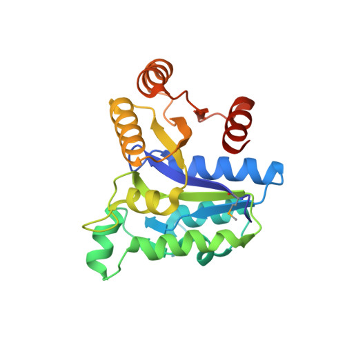 | |
UniProt | |||||
Find proteins for Q5NGB5 (Francisella tularensis subsp. tularensis (strain SCHU S4 / Schu 4)) Explore Q5NGB5 Go to UniProtKB: Q5NGB5 | |||||
Entity Groups | |||||
| Sequence Clusters | 30% Identity50% Identity70% Identity90% Identity95% Identity100% Identity | ||||
| UniProt Group | Q5NGB5 | ||||
Sequence AnnotationsExpand | |||||
| |||||
| Ligands 2 Unique | |||||
|---|---|---|---|---|---|
| ID | Chains | Name / Formula / InChI Key | 2D Diagram | 3D Interactions | |
| ACT Query on ACT | C [auth A], F [auth A], G [auth B] | ACETATE ION C2 H3 O2 QTBSBXVTEAMEQO-UHFFFAOYSA-M |  | ||
| NA Query on NA | D [auth A], E [auth A], H [auth B], I [auth B], J [auth B] | SODIUM ION Na FKNQFGJONOIPTF-UHFFFAOYSA-N |  | ||
| Modified Residues 1 Unique | |||||
|---|---|---|---|---|---|
| ID | Chains | Type | Formula | 2D Diagram | Parent |
| MSE Query on MSE | A, B | L-PEPTIDE LINKING | C5 H11 N O2 Se |  | MET |
| Length ( Å ) | Angle ( ˚ ) |
|---|---|
| a = 39.278 | α = 90 |
| b = 93.177 | β = 95.78 |
| c = 62.732 | γ = 90 |
| Software Name | Purpose |
|---|---|
| BLU-MAX | data collection |
| PHENIX | model building |
| BUSTER | refinement |
| HKL-2000 | data reduction |
| HKL-2000 | data scaling |
| PHENIX | phasing |
RCSB PDB (citation) is hosted by
RCSB PDB is a member of the

