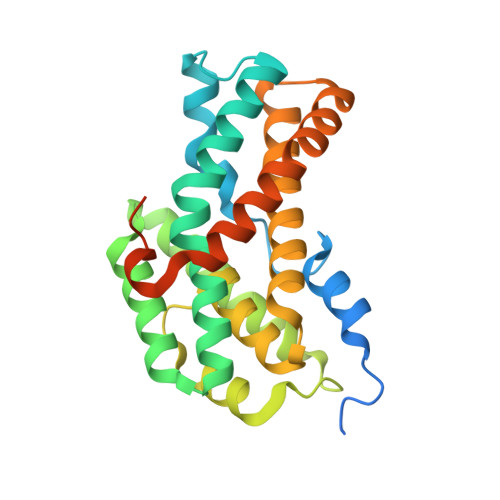
Assembly of Preactivation Complex for Urease Maturation in Helicobacter pylori: CRYSTAL STRUCTURE OF UreF-UreH PROTEIN COMPLEX.
Fong, Y.H., Wong, H.C., Chuck, C.P., Chen, Y.W., Sun, H., Wong, K.B.(2011) J Biol Chem 286: 43241-43249
- PubMed: 22013070
- DOI: https://doi.org/10.1074/jbc.M111.296830
- Primary Citation of Related Structures:
3O1Q, 3SF5 - PubMed Abstract:
Colonization of Helicobacter pylori in the acidic environment of the human stomach depends on the neutralizing activity of urease. Activation of apo-urease involves carboxylation of lysine 219 and insertion of two nickel ions. In H. pylori, this maturation process involves four urease accessory proteins as follows: UreE, UreF, UreG, and UreH. It is postulated that the apo-urease interacts with UreF, UreG, and UreH to form a pre-activation complex that undergoes GTP-dependent activation of urease. The crystal structure of the UreF-UreH complex reveals conformational changes in two distinct regions of UreF upon complex formation. First, the flexible C-terminal residues of UreF become ordered, forming an extra helix α10 and a loop structure stabilized by hydrogen bonds involving Arg-250. Second, the first turn of helix α2 uncoils to expose a conserved residue, Tyr-48. Substitution of R250A or Y48A in UreF abolishes the formation of the heterotrimeric complex of UreG-UreF-UreH and abolishes urease maturation. Our results suggest that the C-terminal residues and helix α2 of UreF are essential for the recruitment of UreG for the formation of the pre-activation complex. The molecular mass of the UreF-UreH complex determined by static light scattering was 116 ± 2.3 kDa, which is consistent with the quaternary structure of a dimer of heterodimers observed in the crystal structure. Taking advantage of the unique 2-fold symmetry observed in both the crystal structures of H. pylori urease and the UreF-UreH complex, we proposed a topology model of the pre-activation complex for urease maturation.
Organizational Affiliation:
Centre for Protein Science and Crystallography, School of Life Sciences, Chinese University of Hong Kong, China, Hong Kong.