3NXN

X-ray structure of ester chemical analogue 'covalent dimer' [Ile50,O-Ile50']HIV-1 protease complexed with KVS-1 inhibitor


Experimental Data Snapshot

  • Method: X-RAY DIFFRACTION
  • Resolution: 1.80 Å
  • R-Value Free: 0.225 
  • R-Value Work: 0.194 
  • R-Value Observed: 0.195 

wwPDB Validation   3D Report Full Report


Ligand Structure Quality Assessment 


This is version 2.0 of the entry. See complete history


Literature

Protein conformational dynamics in the mechanism of HIV-1 protease catalysis.

Torbeev, V.Y.Raghuraman, H.Hamelberg, D.Tonelli, M.Westler, W.M.Perozo, E.Kent, S.B.

(2011) Proc Natl Acad Sci U S A 108: 20982-20987

  • DOI: https://doi.org/10.1073/pnas.1111202108
  • Primary Citation of Related Structures:  
    3FSM, 3HAU, 3HAW, 3HBO, 3HDK, 3HLO, 3IAW, 3KA2, 3NWQ, 3NWX, 3NXE, 3NXN, 3NYG

  • PubMed Abstract: 

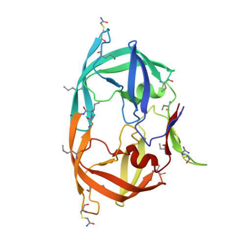
    We have used chemical protein synthesis and advanced physical methods to probe dynamics-function correlations for the HIV-1 protease, an enzyme that has received considerable attention as a target for the treatment of AIDS. Chemical synthesis was used to prepare a series of unique analogues of the HIV-1 protease in which the flexibility of the "flap" structures (residues 37-61 in each monomer of the homodimeric protein molecule) was systematically varied. These analogue enzymes were further studied by X-ray crystallography, NMR relaxation, and pulse-EPR methods, in conjunction with molecular dynamics simulations. We show that conformational isomerization in the flaps is correlated with structural reorganization of residues in the active site, and that it is preorganization of the active site that is a rate-limiting factor in catalysis.


  • Organizational Affiliation

    Department of Chemistry, Institute for Biophysical Dynamics, University of Chicago, Chicago, IL 60637, USA.


Macromolecules
Find similar proteins by:  (by identity cutoff)  |  3D Structure
Entity ID: 1
MoleculeChains Sequence LengthOrganismDetailsImage
protease covalent dimer203N/AMutation(s): 0 
EC: 3.4.23.16
UniProt
Find proteins for P03369 (Human immunodeficiency virus type 1 group M subtype B (isolate ARV2/SF2))
Explore P03369 
Go to UniProtKB:  P03369
Entity Groups  
Sequence Clusters30% Identity50% Identity70% Identity90% Identity95% Identity100% Identity
UniProt GroupP03369
Sequence Annotations
Expand
  • Reference Sequence
Small Molecules
Modified Residues  3 Unique
IDChains TypeFormula2D DiagramParent
ABA
Query on ABA
A
L-PEPTIDE LINKINGC4 H9 N O2ALA
NLE
Query on NLE
A
L-PEPTIDE LINKINGC6 H13 N O2LEU
YCM
Query on YCM
A
L-PEPTIDE LINKINGC5 H10 N2 O3 SCYS
Experimental Data & Validation

Experimental Data

  • Method: X-RAY DIFFRACTION
  • Resolution: 1.80 Å
  • R-Value Free: 0.225 
  • R-Value Work: 0.194 
  • R-Value Observed: 0.195 
  • Space Group: P 21 21 21
Unit Cell:
Length ( Å )Angle ( ˚ )
a = 51.871α = 90
b = 57.644β = 90
c = 61.64γ = 90
Software Package:
Software NamePurpose
ADSCdata collection
MOLREPphasing
REFMACrefinement
HKL-2000data reduction
HKL-2000data scaling

Structure Validation

View Full Validation Report


Ligand Structure Quality Assessment 


Entry History 

Deposition Data

Revision History  (Full details and data files)

  • Version 1.0: 2011-11-02
    Type: Initial release
  • Version 1.1: 2012-01-11
    Changes: Database references
  • Version 1.2: 2012-12-12
    Changes: Other
  • Version 2.0: 2019-07-31
    Changes: Data collection, Derived calculations, Non-polymer description, Structure summary