3NX5

The crystal structure of Sanguinarine bound to DNA d(CGTACG)


Experimental Data Snapshot

  • Method: X-RAY DIFFRACTION
  • Resolution: 2.31 Å
  • R-Value Free: 0.283 
  • R-Value Work: 0.252 
  • R-Value Observed: 0.255 

wwPDB Validation   3D Report Full Report


Ligand Structure Quality Assessment 


This is version 1.2 of the entry. See complete history


Literature

X-Ray diffraction analyses of the natural isoquinoline alkaloids Berberine and Sanguinarine complexed with double helix DNA d(CGTACG)

Ferraroni, M.Bazzicalupi, C.Bilia, A.R.Gratteri, P.

(2011) Chem Commun (Camb) 47: 4917-4919

  • DOI: https://doi.org/10.1039/c1cc10971e
  • Primary Citation of Related Structures:  
    3NP6, 3NX5

  • PubMed Abstract: 

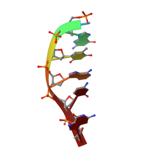
    The first crystal structures of Berberine and Sanguinarine intercalated with a d(CGTACG)(2) DNA sequence were obtained by X-ray diffraction analysis at 2.3 Å resolution. Both drugs join the end of two "two-molecules" DNA units, stacked in a non-classic intercalation site formed by six bases. Sanguinarine interacts with d(CGTACG)(2) DNA in its iminium form.


  • Organizational Affiliation

    Department of Chemistry Ugo Schiff, University of Firenze, Via della Lastruccia 3-13, I-50019 Sesto Fiorentino, Firenze, Italy.


Macromolecules

Find similar nucleic acids by:  Sequence   |   3D Structure  

Entity ID: 1
MoleculeChains LengthOrganismImage
5'-D(*CP*GP*TP*AP*CP*G)-3'
A, B, C, D
6N/A
Sequence Annotations
Expand
  • Reference Sequence
Small Molecules
Ligands 2 Unique
IDChains Name / Formula / InChI Key2D Diagram3D Interactions
SAU
Query on SAU

Download Ideal Coordinates CCD File 
E [auth C]13-methyl[1,3]benzodioxolo[5,6-c][1,3]dioxolo[4,5-i]phenanthridin-13-ium
C20 H14 N O4
INVGWHRKADIJHF-UHFFFAOYSA-N
CA
Query on CA

Download Ideal Coordinates CCD File 
F [auth D]CALCIUM ION
Ca
BHPQYMZQTOCNFJ-UHFFFAOYSA-N
Experimental Data & Validation

Experimental Data

  • Method: X-RAY DIFFRACTION
  • Resolution: 2.31 Å
  • R-Value Free: 0.283 
  • R-Value Work: 0.252 
  • R-Value Observed: 0.255 
  • Space Group: P 32 2 1
Unit Cell:
Length ( Å )Angle ( ˚ )
a = 29.992α = 90
b = 29.992β = 90
c = 119.069γ = 120
Software Package:
Software NamePurpose
XSCALEdata scaling
MOLREPphasing
REFMACrefinement
PDB_EXTRACTdata extraction
XDSdata reduction
XDSdata scaling

Structure Validation

View Full Validation Report


Ligand Structure Quality Assessment 


Entry History 

Deposition Data

Revision History  (Full details and data files)

  • Version 1.0: 2011-05-04
    Type: Initial release
  • Version 1.1: 2011-07-13
    Changes: Version format compliance
  • Version 1.2: 2023-11-01
    Changes: Data collection, Database references, Derived calculations, Refinement description