


Crystal structure of the first omega-transaminase at 2.0A resolution
Park, H.H., Jang, T.To be published.
Experimental Data Snapshot
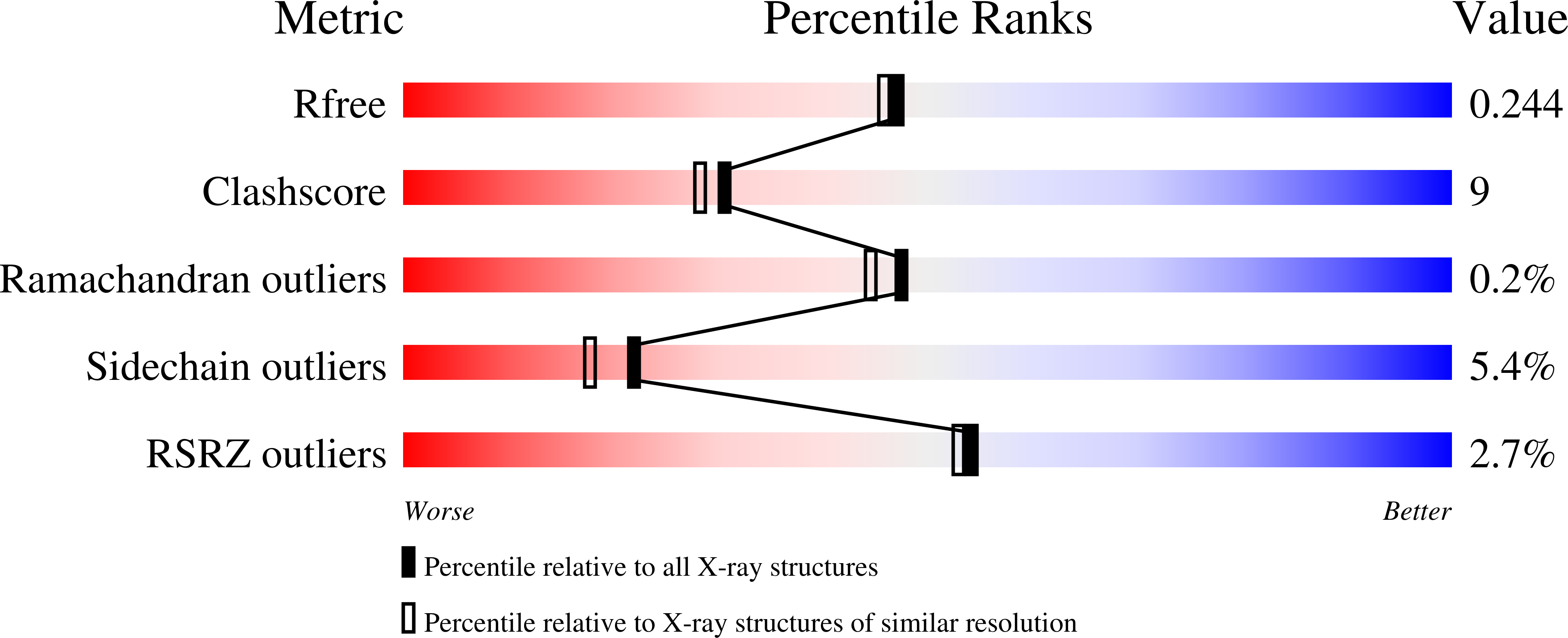
wwPDB Validation 3D Report Full Report
Entity ID: 1 | |||||
|---|---|---|---|---|---|
| Molecule | Chains | Sequence Length | Organism | Details | Image |
| Pyruvate transaminase | 478 | Vibrio fluvialis | Mutation(s): 0 | 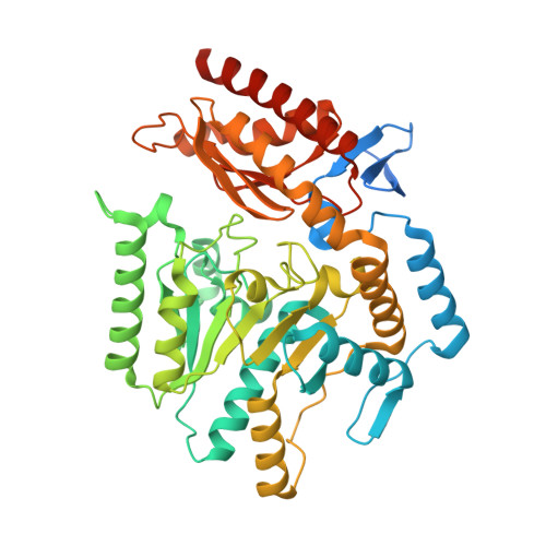 | |
UniProt | |||||
Find proteins for F2XBU9 (Vibrio fluvialis) Explore F2XBU9 Go to UniProtKB: F2XBU9 | |||||
Entity Groups | |||||
| Sequence Clusters | 30% Identity50% Identity70% Identity90% Identity95% Identity100% Identity | ||||
| UniProt Group | F2XBU9 | ||||
Sequence AnnotationsExpand | |||||
| |||||
| Length ( Å ) | Angle ( ˚ ) |
|---|---|
| a = 78.075 | α = 90 |
| b = 95.03 | β = 90 |
| c = 123.079 | γ = 90 |
| Software Name | Purpose |
|---|---|
| REFMAC | refinement |
RCSB PDB (citation) is hosted by
RCSB PDB is a member of the

