


Crystal Structure of Human Peripheral Myelin Protein 2
Ugochukwu, E., Pilka, E., Phillips, C., Yue, W.W., Krojer, T., von Delft, F., Bountra, C., Arrowsmith, C.H., Weigelt, J., Edwards, A., Kavanagh, K.L., Oppermann, U.To be published.
Experimental Data Snapshot
Entity ID: 1 | |||||
|---|---|---|---|---|---|
| Molecule | Chains | Sequence Length | Organism | Details | Image |
| PMP2 protein | 153 | Homo sapiens | Mutation(s): 0 Gene Names: hCG_21300, PMP2 | 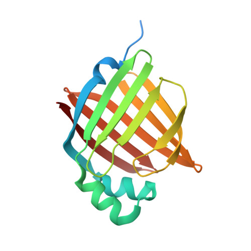 | |
UniProt & NIH Common Fund Data Resources | |||||
Find proteins for P02689 (Homo sapiens) Explore P02689 Go to UniProtKB: P02689 | |||||
PHAROS: P02689 GTEx: ENSG00000147588 | |||||
Entity Groups | |||||
| Sequence Clusters | 30% Identity50% Identity70% Identity90% Identity95% Identity100% Identity | ||||
| UniProt Group | P02689 | ||||
Sequence AnnotationsExpand | |||||
| |||||
| Ligands 6 Unique | |||||
|---|---|---|---|---|---|
| ID | Chains | Name / Formula / InChI Key | 2D Diagram | 3D Interactions | |
| PLM Query on PLM | B [auth A] | PALMITIC ACID C16 H32 O2 IPCSVZSSVZVIGE-UHFFFAOYSA-N |  | ||
| EPE Query on EPE | H [auth A] | 4-(2-HYDROXYETHYL)-1-PIPERAZINE ETHANESULFONIC ACID C8 H18 N2 O4 S JKMHFZQWWAIEOD-UHFFFAOYSA-N |  | ||
| CIT Query on CIT | G [auth A] | CITRIC ACID C6 H8 O7 KRKNYBCHXYNGOX-UHFFFAOYSA-N |  | ||
| SO4 Query on SO4 | C [auth A], D [auth A], E [auth A] | SULFATE ION O4 S QAOWNCQODCNURD-UHFFFAOYSA-L |  | ||
| GOL Query on GOL | F [auth A] | GLYCEROL C3 H8 O3 PEDCQBHIVMGVHV-UHFFFAOYSA-N |  | ||
| CL Query on CL | I [auth A] | CHLORIDE ION Cl VEXZGXHMUGYJMC-UHFFFAOYSA-M |  | ||
| Length ( Å ) | Angle ( ˚ ) |
|---|---|
| a = 67.19 | α = 90 |
| b = 67.19 | β = 90 |
| c = 100.07 | γ = 90 |
| Software Name | Purpose |
|---|---|
| MAR345 | data collection |
| PHASER | phasing |
| REFMAC | refinement |
| MOSFLM | data reduction |
| SCALA | data scaling |
RCSB PDB (citation) is hosted by
RCSB PDB is a member of the

