3NBX

Crystal structure of E. coli RavA (Regulatory ATPase variant A) in complex with ADP


Experimental Data Snapshot

  • Method: X-RAY DIFFRACTION
  • Resolution: 2.91 Å
  • R-Value Free: 0.269 
  • R-Value Work: 0.223 
  • R-Value Observed: 0.226 

wwPDB Validation   3D Report Full Report


Ligand Structure Quality Assessment 


This is version 1.3 of the entry. See complete history


Literature

Structure of RavA MoxR AAA+ protein reveals the design principles of a molecular cage modulating the inducible lysine decarboxylase activity

El Bakkouri, M.Gutsche, I.Kanjee, U.Zhao, B.Yu, M.Goret, G.Schoehn, G.Burmeister, W.P.Houry, W.A.

(2010) Proc Natl Acad Sci U S A 107: 22499-22504

  • DOI: https://doi.org/10.1073/pnas.1009092107
  • Primary Citation of Related Structures:  
    3NBX

  • PubMed Abstract: 

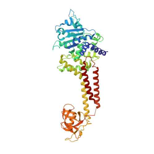
    The MoxR family of AAA+ ATPases is widespread throughout bacteria and archaea but remains poorly characterized. We recently found that the Escherichia coli MoxR protein, RavA (Regulatory ATPase variant A), tightly interacts with the inducible lysine decarboxylase, LdcI/CadA, to form a unique cage-like structure. Here, we present the X-ray structure of RavA and show that the αβα and all-α subdomains in the RavA AAA+ module are arranged as in magnesium chelatases rather than as in classical AAA+ proteins. RavA structure also contains a discontinuous triple-helical domain as well as a β-barrel-like domain forming a unique fold, which we termed the LARA domain. The LARA domain was found to mediate the interaction between RavA and LdcI. The RavA structure provides insights into how five RavA hexamers interact with two LdcI decamers to form the RavA-LdcI cage-like structure.


  • Organizational Affiliation

    Department of Biochemistry, University of Toronto, Toronto, ON, Canada M5S 1A8.


Macromolecules
Find similar proteins by:  (by identity cutoff)  |  3D Structure
Entity ID: 1
MoleculeChains Sequence LengthOrganismDetailsImage
ATPase ravAA [auth X]500Escherichia coli K-12Mutation(s): 0 
Gene Names: b3746JW3725ravAyieN
EC: 3.6.3
UniProt
Find proteins for P31473 (Escherichia coli (strain K12))
Explore P31473 
Go to UniProtKB:  P31473
Entity Groups  
Sequence Clusters30% Identity50% Identity70% Identity90% Identity95% Identity100% Identity
UniProt GroupP31473
Sequence Annotations
Expand
  • Reference Sequence
Small Molecules
Ligands 2 Unique
IDChains Name / Formula / InChI Key2D Diagram3D Interactions
ADP
Query on ADP

Download Ideal Coordinates CCD File 
B [auth X]ADENOSINE-5'-DIPHOSPHATE
C10 H15 N5 O10 P2
XTWYTFMLZFPYCI-KQYNXXCUSA-N
SO4
Query on SO4

Download Ideal Coordinates CCD File 
C [auth X]SULFATE ION
O4 S
QAOWNCQODCNURD-UHFFFAOYSA-L
Experimental Data & Validation

Experimental Data

  • Method: X-RAY DIFFRACTION
  • Resolution: 2.91 Å
  • R-Value Free: 0.269 
  • R-Value Work: 0.223 
  • R-Value Observed: 0.226 
  • Space Group: P 65
Unit Cell:
Length ( Å )Angle ( ˚ )
a = 162.232α = 90
b = 162.232β = 90
c = 55.316γ = 120
Software Package:
Software NamePurpose
DNAdata collection
SHARPphasing
REFMACrefinement
HKL-2000data reduction
HKL-2000data scaling

Structure Validation

View Full Validation Report


Ligand Structure Quality Assessment 


Entry History 

Deposition Data

Revision History  (Full details and data files)

  • Version 1.0: 2011-01-12
    Type: Initial release
  • Version 1.1: 2011-07-13
    Changes: Version format compliance
  • Version 1.2: 2014-11-12
    Changes: Structure summary
  • Version 1.3: 2024-02-21
    Changes: Data collection, Database references, Derived calculations