3MXE

Crystal structure of HIV-1 protease inhibitor, KC32 complexed with wild-type protease


Experimental Data Snapshot

  • Method: X-RAY DIFFRACTION
  • Resolution: 1.85 Å
  • R-Value Free: 0.223 
  • R-Value Work: 0.180 
  • R-Value Observed: 0.182 

wwPDB Validation   3D Report Full Report


Ligand Structure Quality Assessment 


This is version 1.4 of the entry. See complete history


Literature

Structure-Based Design, Synthesis, and Structure-Activity Relationship Studies of HIV-1 Protease Inhibitors Incorporating Phenyloxazolidinones.

Ali, A.Reddy, G.S.Nalam, M.N.Anjum, S.G.Cao, H.Schiffer, C.A.Rana, T.M.

(2010) J Med Chem 53: 7699-7708

  • DOI: https://doi.org/10.1021/jm1008743
  • Primary Citation of Related Structures:  
    3MXD, 3MXE

  • PubMed Abstract: 

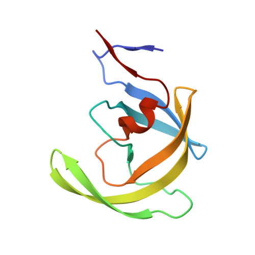
    A series of new HIV-1 protease inhibitors with the hydroxyethylamine core and different phenyloxazolidinone P2 ligands were designed and synthesized. Variation of phenyl substitutions at the P2 and P2' moieties significantly affected the binding affinity and antiviral potency of the inhibitors. In general, compounds with 2- and 4-substituted phenyloxazolidinones at P2 exhibited lower binding affinities than 3-substituted analogues. Crystal structure analyses of ligand-enzyme complexes revealed different binding modes for 2- and 3-substituted P2 moieties in the protease S2 binding pocket, which may explain their different binding affinities. Several compounds with 3-substituted P2 moieties demonstrated picomolar binding affinity and low nanomolar antiviral potency against patient-derived viruses from HIV-1 clades A, B, and C, and most retained potency against drug-resistant viruses. Further optimization of these compounds using structure-based design may lead to the development of novel protease inhibitors with improved activity against drug-resistant strains of HIV-1.


  • Organizational Affiliation

    University of Massachusetts Medical School, Worcester, Massachusetts 01605, United States.


Macromolecules
Find similar proteins by:  (by identity cutoff)  |  3D Structure
Entity ID: 1
MoleculeChains Sequence LengthOrganismDetailsImage
HIV-1 protease
A, B
99HIV-1 M:B_ARV2/SF2Mutation(s): 2 
Gene Names: gag-pol
EC: 3.4.23.16
UniProt
Find proteins for P03369 (Human immunodeficiency virus type 1 group M subtype B (isolate ARV2/SF2))
Explore P03369 
Go to UniProtKB:  P03369
Entity Groups  
Sequence Clusters30% Identity50% Identity70% Identity90% Identity95% Identity100% Identity
UniProt GroupP03369
Sequence Annotations
Expand
  • Reference Sequence
Small Molecules
Ligands 3 Unique
IDChains Name / Formula / InChI Key2D Diagram3D Interactions
K54
Query on K54

Download Ideal Coordinates CCD File 
C [auth A](5S)-N-{(1S,2R)-3-[(1,3-benzothiazol-6-ylsulfonyl)(2-methylpropyl)amino]-1-benzyl-2-hydroxypropyl}-2-oxo-3-[2-(trifluoromethyl)phenyl]-1,3-oxazolidine-5-carboxamide
C32 H33 F3 N4 O6 S2
NMADTNHSJGJGDP-ITHJLQSDSA-N
PO4
Query on PO4

Download Ideal Coordinates CCD File 
D [auth B]PHOSPHATE ION
O4 P
NBIIXXVUZAFLBC-UHFFFAOYSA-K
ACT
Query on ACT

Download Ideal Coordinates CCD File 
E [auth B]ACETATE ION
C2 H3 O2
QTBSBXVTEAMEQO-UHFFFAOYSA-M
Binding Affinity Annotations 
IDSourceBinding Affinity
K54 PDBBind:  3MXE Ki: 0.1 (nM) from 1 assay(s)
Binding MOAD:  3MXE Ki: 0.1 (nM) from 1 assay(s)
BindingDB:  3MXE Ki: 0.1 (nM) from 1 assay(s)
Experimental Data & Validation

Experimental Data

  • Method: X-RAY DIFFRACTION
  • Resolution: 1.85 Å
  • R-Value Free: 0.223 
  • R-Value Work: 0.180 
  • R-Value Observed: 0.182 
  • Space Group: P 21 21 21
Unit Cell:
Length ( Å )Angle ( ˚ )
a = 50.79α = 90
b = 58.245β = 90
c = 62.014γ = 90
Software Package:
Software NamePurpose
REFMACrefinement
DENZOdata reduction
SCALEPACKdata scaling
AMoREphasing

Structure Validation

View Full Validation Report


Ligand Structure Quality Assessment 


Entry History 

Deposition Data

Revision History  (Full details and data files)

  • Version 1.0: 2010-11-10
    Type: Initial release
  • Version 1.1: 2011-07-13
    Changes: Version format compliance
  • Version 1.2: 2017-11-08
    Changes: Refinement description
  • Version 1.3: 2023-09-06
    Changes: Data collection, Database references, Derived calculations, Refinement description
  • Version 1.4: 2024-03-13
    Changes: Source and taxonomy, Structure summary