3MQY

SgrAI with cleaved DNA and Magnesium bound


Experimental Data Snapshot

  • Method: X-RAY DIFFRACTION
  • Resolution: 2.00 Å
  • R-Value Free: 0.249 
  • R-Value Work: 0.196 
  • R-Value Observed: 0.199 

Starting Model: experimental
View more details

wwPDB Validation   3D Report Full Report


This is version 1.2 of the entry. See complete history


Literature

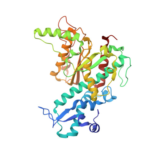
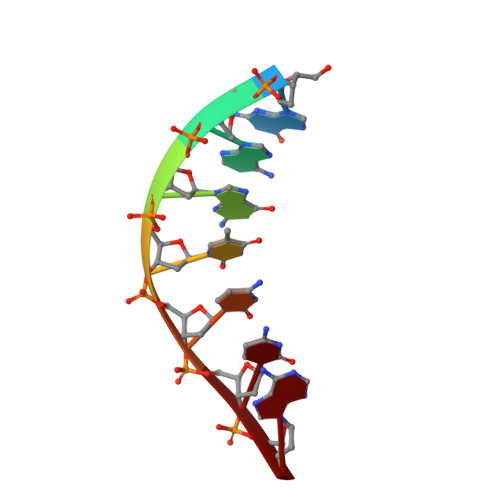
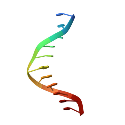
New clues in the allosteric activation of DNA cleavage by SgrAI: structures of SgrAI bound to cleaved primary-site DNA and uncleaved secondary-site DNA.

Little, E.J.Dunten, P.W.Bitinaite, J.Horton, N.C.

(2011) Acta Crystallogr D Biol Crystallogr 67: 67-74

  • DOI: https://doi.org/10.1107/S0907444910047785
  • Primary Citation of Related Structures:  
    3MQY, 3N78, 3N7B

  • PubMed Abstract: 

    SgrAI is a type II restriction endonuclease that cuts an unusually long recognition sequence and exhibits allosteric self-activation with expansion of DNA-sequence specificity. The three-dimensional crystal structures of SgrAI bound to cleaved primary-site DNA and Mg²(+) and bound to secondary-site DNA with either Mg²(+) or Ca²(+) are presented. All three structures show a conformation of enzyme and DNA similar to the previously determined dimeric structure of SgrAI bound to uncleaved primary-site DNA and Ca²(+) [Dunten et al. (2008), Nucleic Acids Res. 36, 5405-5416], with the exception of the cleaved bond and a slight shifting of the DNA in the SgrAI/cleaved primary-site DNA/Mg²(+) structure. In addition, a new metal ion binding site is located in one of the two active sites in this structure, which is consistent with proposals for the existence of a metal-ion site near the 3'-O leaving group.


  • Organizational Affiliation

    Department of Chemistry and Biochemistry, University of Arizona, Tucson, 85721, USA.


Macromolecules

Find similar proteins by:  (by identity cutoff)  |  3D Structure
Entity ID: 1
MoleculeChains Sequence LengthOrganismDetailsImage
SgraIR restriction enzyme
A, B
338Streptomyces griseusMutation(s): 1 
Gene Names: sgraIR
EC: 3.1.21.4
UniProt
Find proteins for Q9F6L0 (Streptomyces griseus)
Explore Q9F6L0 
Go to UniProtKB:  Q9F6L0
Entity Groups  
Sequence Clusters30% Identity50% Identity70% Identity90% Identity95% Identity100% Identity
UniProt GroupQ9F6L0
Sequence Annotations
Expand
  • Reference Sequence

Find similar nucleic acids by:  Sequence   |   3D Structure  

Entity ID: 2
MoleculeChains LengthOrganismImage
DNA (5'-D(*GP*AP*GP*TP*CP*CP*A)-3')
C, E
7N/A
Sequence Annotations
Expand
  • Reference Sequence

Find similar nucleic acids by:  Sequence   |   3D Structure  

Entity ID: 3
MoleculeChains LengthOrganismImage
DNA (5'-D(P*CP*CP*GP*GP*TP*GP*GP*AP*CP*TP*C)-3')
D, F
11N/A
Sequence Annotations
Expand
  • Reference Sequence
Experimental Data & Validation

Experimental Data

  • Method: X-RAY DIFFRACTION
  • Resolution: 2.00 Å
  • R-Value Free: 0.249 
  • R-Value Work: 0.196 
  • R-Value Observed: 0.199 
  • Space Group: P 1 21 1
Unit Cell:
Length ( Å )Angle ( ˚ )
a = 59.163α = 90
b = 119.98β = 102.68
c = 69.857γ = 90
Software Package:
Software NamePurpose
Blu-Icedata collection
MOLREPphasing
REFMACrefinement
XDSdata reduction
XSCALEdata scaling

Structure Validation

View Full Validation Report


Entry History 

Deposition Data

Revision History  (Full details and data files)

  • Version 1.0: 2010-11-24
    Type: Initial release
  • Version 1.1: 2011-07-13
    Changes: Version format compliance
  • Version 1.2: 2023-09-06
    Changes: Data collection, Database references, Derived calculations, Refinement description