3MMI

Crystal structure of the globular tail of Myo4p


Experimental Data Snapshot

  • Method: X-RAY DIFFRACTION
  • Resolution: 2.30 Å
  • R-Value Free: 0.287 
  • R-Value Work: 0.254 
  • R-Value Observed: 0.254 

wwPDB Validation   3D Report Full Report


This is version 1.2 of the entry. See complete history


Literature

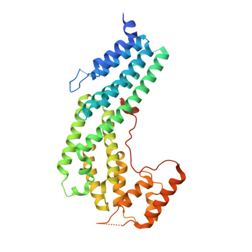
The structure of the Myo4p globular tail and its function in ASH1 mRNA localization.

Heuck, A.Fetka, I.Brewer, D.N.Huls, D.Munson, M.Jansen, R.P.Niessing, D.

(2010) J Cell Biol 189: 497-510

  • DOI: https://doi.org/10.1083/jcb.201002076
  • Primary Citation of Related Structures:  
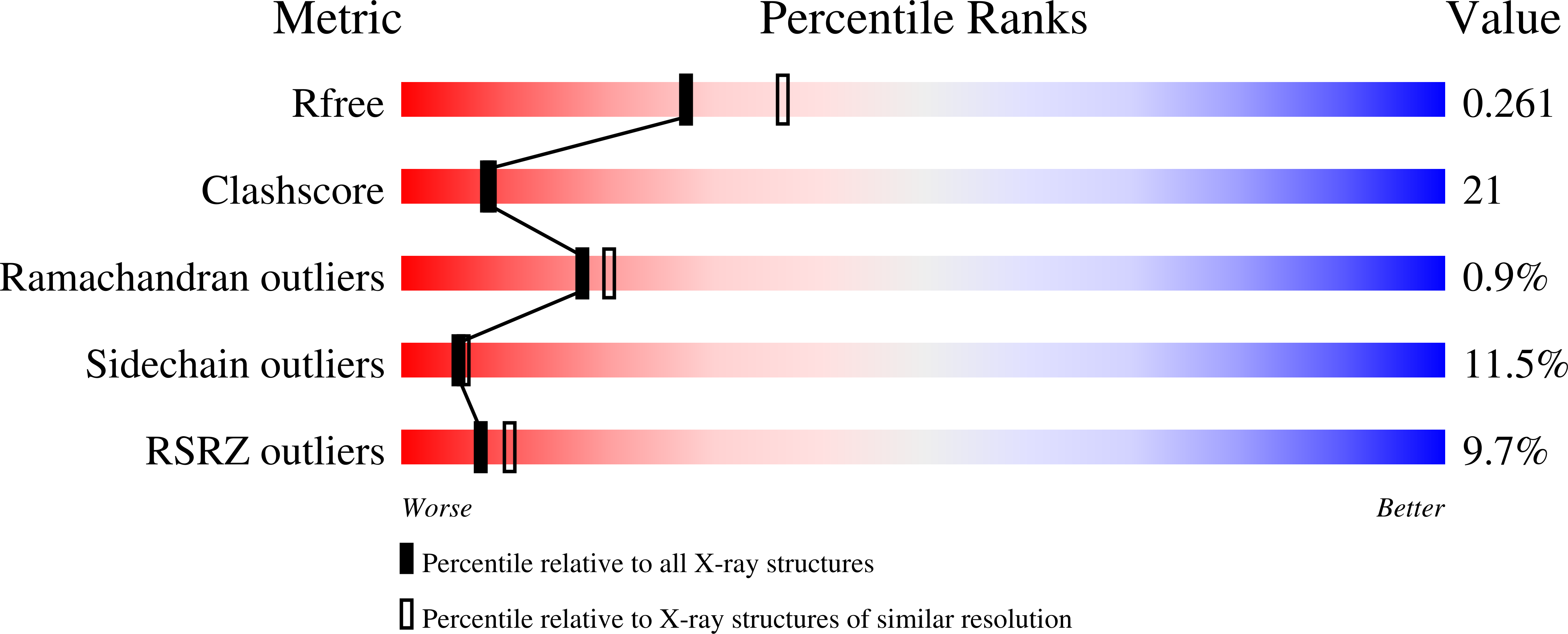
    3MMI

  • PubMed Abstract: 

    Type V myosin (MyoV)-dependent transport of cargo is an essential process in eukaryotes. Studies on yeast and vertebrate MyoV showed that their globular tails mediate binding to the cargo complexes. In Saccharomyces cerevisiae, the MyoV motor Myo4p interacts with She3p to localize asymmetric synthesis of HO 1 (ASH1) mRNA into the bud of dividing cells. A recent study showed that localization of GFP-MS2-tethered ASH1 particles does not require the Myo4p globular tail, challenging the supposed role of this domain. We assessed ASH1 mRNA and Myo4p distribution more directly and found that their localization is impaired in cells expressing globular tail-lacking Myo4p. In vitro studies further show that the globular tail together with a more N-terminal linker region is required for efficient She3p binding. We also determined the x-ray structure of the Myo4p globular tail and identify a conserved surface patch important for She3p binding. The structure shows pronounced similarities to membrane-tethering complexes and indicates that Myo4p may not undergo auto-inhibition of its motor domain.


  • Organizational Affiliation

    Institute of Structural Biology, Helmholtz Zentrum München, German Research Center for Environmental Health, Munich, Germany.


Macromolecules
Find similar proteins by:  (by identity cutoff)  |  3D Structure
Entity ID: 1
MoleculeChains Sequence LengthOrganismDetailsImage
Myosin-4
A, B
386Saccharomyces cerevisiaeMutation(s): 0 
Gene Names: FUN22MYO4SHE1YAL029C
UniProt
Find proteins for P32492 (Saccharomyces cerevisiae (strain ATCC 204508 / S288c))
Explore P32492 
Go to UniProtKB:  P32492
Entity Groups  
Sequence Clusters30% Identity50% Identity70% Identity90% Identity95% Identity100% Identity
UniProt GroupP32492
Sequence Annotations
Expand
  • Reference Sequence
Experimental Data & Validation

Experimental Data

  • Method: X-RAY DIFFRACTION
  • Resolution: 2.30 Å
  • R-Value Free: 0.287 
  • R-Value Work: 0.254 
  • R-Value Observed: 0.254 
  • Space Group: P 21 21 21
Unit Cell:
Length ( Å )Angle ( ˚ )
a = 43.476α = 90
b = 120.989β = 90
c = 157.683γ = 90
Software Package:
Software NamePurpose
ADSCdata collection
SHELXDphasing
REFMACrefinement
MOSFLMdata reduction
SCALAdata scaling

Structure Validation

View Full Validation Report


Entry History 

Deposition Data

Revision History  (Full details and data files)

  • Version 1.0: 2010-05-12
    Type: Initial release
  • Version 1.1: 2011-07-13
    Changes: Version format compliance
  • Version 1.2: 2024-02-21
    Changes: Data collection, Database references