3MAZ

Crystal Structure of the Human BRDG1/STAP-1 SH2 Domain in Complex with the NTAL pTyr136 Peptide


Experimental Data Snapshot

  • Method: X-RAY DIFFRACTION
  • Resolution: 1.90 Å
  • R-Value Free: 0.236 
  • R-Value Work: 0.212 
  • R-Value Observed: 0.212 

wwPDB Validation   3D Report Full Report


This is version 1.4 of the entry. See complete history


Literature

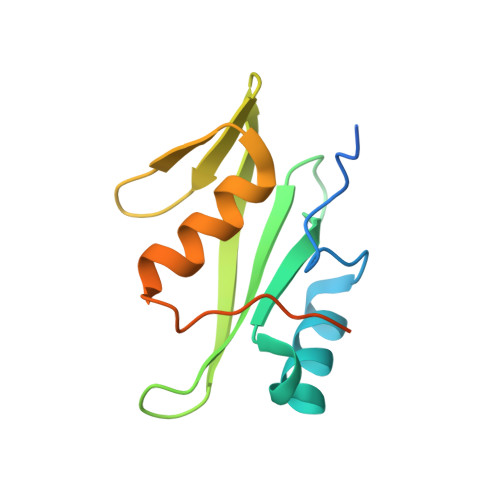
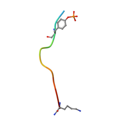
Loops govern SH2 domain specificity by controlling access to binding pockets.

Kaneko, T.Huang, H.Zhao, B.Li, L.Liu, H.Voss, C.K.Wu, C.Schiller, M.R.Li, S.S.

(2010) Sci Signal 3: ra34-ra34

  • DOI: https://doi.org/10.1126/scisignal.2000796
  • Primary Citation of Related Structures:  
    3MAZ

  • PubMed Abstract: 

    Cellular functions require specific protein-protein interactions that are often mediated by modular domains that use binding pockets to engage particular sequence motifs in their partners. Yet, how different members of a domain family select for distinct sequence motifs is not fully understood. The human genome encodes 120 Src homology 2 (SH2) domains (in 110 proteins), which mediate protein-protein interactions by binding to proteins with diverse phosphotyrosine (pTyr)-containing sequences. The structure of the SH2 domain of BRDG1 bound to a peptide revealed a binding pocket that was blocked by a loop residue in most other SH2 domains. Analysis of 63 SH2 domain structures suggested that the SH2 domains contain three binding pockets, which exhibit selectivity for the three positions after the pTyr in a peptide, and that SH2 domain loops defined the accessibility and shape of these pockets. Despite sequence variability in the loops, we identified conserved structural features in the loops of SH2 domains responsible for controlling access to these surface pockets. We engineered new loops in an SH2 domain that altered specificity as predicted. Thus, selective blockage of binding subsites or pockets by surface loops provides a molecular basis by which the diverse modes of ligand recognition by the SH2 domain may have evolved and provides a framework for engineering SH2 domains and designing SH2-specific inhibitors.


  • Organizational Affiliation

    Department of Biochemistry and the Siebens-Drake Research Institute, Schulich School of Medicine and Dentistry, University of Western Ontario, London, Ontario, Canada N6A 5C1.


Macromolecules
Find similar proteins by:  (by identity cutoff)  |  3D Structure
Entity ID: 1
MoleculeChains Sequence LengthOrganismDetailsImage
Signal-transducing adaptor protein 1125Homo sapiensMutation(s): 1 
Gene Names: STAP1BRDG1
UniProt & NIH Common Fund Data Resources
Find proteins for Q9ULZ2 (Homo sapiens)
Explore Q9ULZ2 
Go to UniProtKB:  Q9ULZ2
PHAROS:  Q9ULZ2
GTEx:  ENSG00000035720 
Entity Groups  
Sequence Clusters30% Identity50% Identity70% Identity90% Identity95% Identity100% Identity
UniProt GroupQ9ULZ2
Sequence Annotations
Expand
  • Reference Sequence

Find similar proteins by:  Sequence   |   3D Structure  

Entity ID: 2
MoleculeChains Sequence LengthOrganismDetailsImage
CheD family protein12N/AMutation(s): 2 
UniProt & NIH Common Fund Data Resources
Find proteins for Q9GZY6 (Homo sapiens)
Explore Q9GZY6 
Go to UniProtKB:  Q9GZY6
PHAROS:  Q9GZY6
GTEx:  ENSG00000086730 
Entity Groups  
Sequence Clusters30% Identity50% Identity70% Identity90% Identity95% Identity100% Identity
UniProt GroupQ9GZY6
Sequence Annotations
Expand
  • Reference Sequence
Small Molecules
Ligands 1 Unique
IDChains Name / Formula / InChI Key2D Diagram3D Interactions
MLI
Query on MLI

Download Ideal Coordinates CCD File 
C [auth A]MALONATE ION
C3 H2 O4
OFOBLEOULBTSOW-UHFFFAOYSA-L
Modified Residues  1 Unique
IDChains TypeFormula2D DiagramParent
PTR
Query on PTR
B
L-PEPTIDE LINKINGC9 H12 N O6 PTYR
Experimental Data & Validation

Experimental Data

  • Method: X-RAY DIFFRACTION
  • Resolution: 1.90 Å
  • R-Value Free: 0.236 
  • R-Value Work: 0.212 
  • R-Value Observed: 0.212 
  • Space Group: P 62 2 2
Unit Cell:
Length ( Å )Angle ( ˚ )
a = 72.64α = 90
b = 72.64β = 90
c = 97.84γ = 120
Software Package:
Software NamePurpose
CrystalCleardata collection
PHASERphasing
CNSrefinement
CrystalCleardata reduction
CrystalCleardata scaling

Structure Validation

View Full Validation Report


Entry History 

Deposition Data

Revision History  (Full details and data files)

  • Version 1.0: 2010-05-12
    Type: Initial release
  • Version 1.1: 2011-07-13
    Changes: Version format compliance
  • Version 1.2: 2021-10-06
    Changes: Database references, Derived calculations
  • Version 1.3: 2023-09-06
    Changes: Data collection, Refinement description
  • Version 1.4: 2023-11-22
    Changes: Data collection