3M6S

Crystal structure of H1N1pdm Hemagglutinin


Experimental Data Snapshot

  • Method: X-RAY DIFFRACTION
  • Resolution: 2.80 Å
  • R-Value Free: 0.256 
  • R-Value Work: 0.230 
  • R-Value Observed: 0.231 

wwPDB Validation   3D Report Full Report


Ligand Structure Quality Assessment 


This is version 2.2 of the entry. See complete history


Literature

Structure and Receptor binding properties of a pandemic H1N1 virus hemagglutinin

Yang, H.Carney, P.Stevens, J.

(2010) PLoS Curr : RRN1152-RRN1152

  • DOI: https://doi.org/10.1371/currents.RRN1152
  • Primary Citation of Related Structures:  
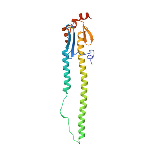
    3M6S

  • PubMed Abstract: 

    The 3D-structure of the major surface viral antigen from the recent H1N1 pandemic influenza virus (A/Darwin/2001/2009) was determined to 2.8 Å resolution. The structure was used to analyze changes in the HA that have emerged during the first 11 months of the pandemic and have raised public health concerns. Receptor binding properties of this protein reveals a strict preference for human-type receptors.


  • Organizational Affiliation

    Centers for Disease Control and Prevention.


Macromolecules
Find similar proteins by:  (by identity cutoff)  |  3D Structure
Entity ID: 1
MoleculeChains Sequence LengthOrganismDetailsImage
Hemagglutinin
A, C, E, G, I
A, C, E, G, I, K
331Influenza A virus (A/Darwin/2001/2009(H1N1))Mutation(s): 0 
Gene Names: HA
UniProt
Find proteins for C5MV42 (Influenza A virus)
Explore C5MV42 
Go to UniProtKB:  C5MV42
Entity Groups  
Sequence Clusters30% Identity50% Identity70% Identity90% Identity95% Identity100% Identity
UniProt GroupC5MV42
Sequence Annotations
Expand
  • Reference Sequence
Find similar proteins by:  (by identity cutoff)  |  3D Structure
Entity ID: 2
MoleculeChains Sequence LengthOrganismDetailsImage
Hemagglutinin
B, D, F, H, J
B, D, F, H, J, L
181Influenza A virus (A/Darwin/2001/2009(H1N1))Mutation(s): 3 
Gene Names: HA
UniProt
Find proteins for C5MV42 (Influenza A virus)
Explore C5MV42 
Go to UniProtKB:  C5MV42
Entity Groups  
Sequence Clusters30% Identity50% Identity70% Identity90% Identity95% Identity100% Identity
UniProt GroupC5MV42
Sequence Annotations
Expand
  • Reference Sequence
Oligosaccharides

Help

Entity ID: 3
MoleculeChains Length2D Diagram Glycosylation3D Interactions
2-acetamido-2-deoxy-beta-D-glucopyranose-(1-4)-2-acetamido-2-deoxy-beta-D-glucopyranose
M, O, P, Q
2N-Glycosylation
Glycosylation Resources
GlyTouCan:  G42666HT
GlyCosmos:  G42666HT
GlyGen:  G42666HT
Entity ID: 4
MoleculeChains Length2D Diagram Glycosylation3D Interactions
alpha-D-mannopyranose-(1-4)-2-acetamido-2-deoxy-beta-D-glucopyranose-(1-4)-2-acetamido-2-deoxy-beta-D-glucopyranose
N
3N-Glycosylation
Glycosylation Resources
GlyTouCan:  G62182OO
GlyCosmos:  G62182OO
GlyGen:  G62182OO
Experimental Data & Validation

Experimental Data

  • Method: X-RAY DIFFRACTION
  • Resolution: 2.80 Å
  • R-Value Free: 0.256 
  • R-Value Work: 0.230 
  • R-Value Observed: 0.231 
  • Space Group: P 1
Unit Cell:
Length ( Å )Angle ( ˚ )
a = 73.984α = 86.25
b = 109.709β = 74.68
c = 129.896γ = 75.1
Software Package:
Software NamePurpose
HKL-2000data collection
PHASERphasing
REFMACrefinement
HKL-2000data reduction
HKL-2000data scaling

Structure Validation

View Full Validation Report


Ligand Structure Quality Assessment 


Entry History 

Deposition Data

Revision History  (Full details and data files)

  • Version 1.0: 2010-03-31
    Type: Initial release
  • Version 1.1: 2011-07-13
    Changes: Advisory, Refinement description, Version format compliance
  • Version 1.2: 2014-03-19
    Changes: Database references
  • Version 2.0: 2020-07-29
    Type: Remediation
    Reason: Carbohydrate remediation
    Changes: Advisory, Atomic model, Data collection, Database references, Derived calculations, Structure summary
  • Version 2.1: 2021-10-06
    Changes: Advisory, Database references, Structure summary
  • Version 2.2: 2023-09-06
    Changes: Data collection, Refinement description