3LLL

Crystal structure of mouse pacsin2 F-BAR domain


Experimental Data Snapshot

  • Method: X-RAY DIFFRACTION
  • Resolution: 3.30 Å
  • R-Value Free: 0.297 
  • R-Value Work: 0.256 
  • R-Value Observed: 0.258 

wwPDB Validation   3D Report Full Report


This is version 1.2 of the entry. See complete history


Literature

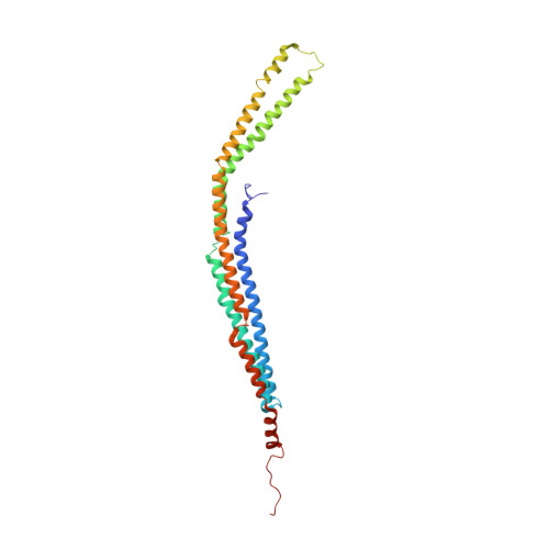
A hinge in the distal end of the PACSIN 2 F-BAR domain may contribute to membrane-curvature sensing.

Plomann, M.Wittmann, J.G.Rudolph, M.G.

(2010) J Mol Biol 400: 129-136

  • DOI: https://doi.org/10.1016/j.jmb.2010.05.008
  • Primary Citation of Related Structures:  
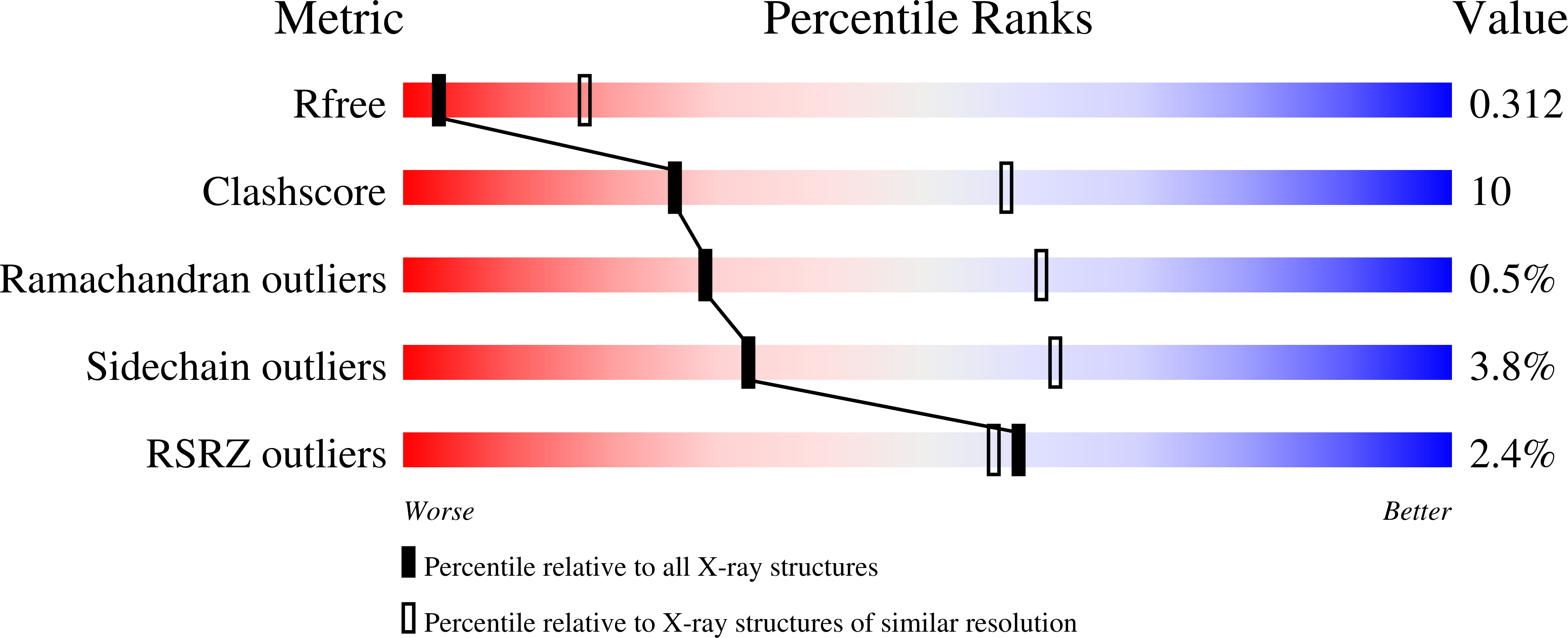
    3LLL

  • PubMed Abstract: 

    The protein kinase C and casein kinase 2 substrates in neurons (PACSINs) represent a subfamily of membrane-binding proteins characterized by an amino-terminal Bin-Amphiphysin-Rvs (F-BAR) domain. PACSINs link membrane trafficking with actin dynamics and regulate the localization of distinct cargo molecules. The F-BAR domain forms a dimer essential for lipid binding. We have obtained crystals of authentic murine PACSIN 2 that contain an ordered F-BAR domain, indicating that additional domains are flexibly connected to F-BAR. The structure shares similarity to other BAR domains and exhibits special features unique to PACSINs. These include the uneven distribution of charged residues on the concave molecular surface and a so-called wedge loop that is driven into the membrane upon binding of PACSIN. The murine PACSIN 2 F-BAR domain requires dimerization for sensing of curved membranes, and the present structure also provides a mechanism for higher-order oligomer formation. Importantly, comparison of murine with human and Drosophila PACSIN 2 F-BAR domains reveals stark differences in the orientation of distal helical segments leading to a wider crescent shape of murine PACSIN 2. We define hinge residues for these movements that may help PACSINs sense and concomitantly reinforce membrane curvature.


  • Organizational Affiliation

    Institute for Biochemistry II and Center for Molecular Medicine Cologne (CMMC), University of Cologne, D-50931 Cologne, Germany.


Macromolecules
Find similar proteins by:  (by identity cutoff)  |  3D Structure
Entity ID: 1
MoleculeChains Sequence LengthOrganismDetailsImage
Protein kinase C and casein kinase substrate in neurons protein 2
A, B
287Mus musculusMutation(s): 0 
Gene Names: Pacsin2
Membrane Entity: Yes 
UniProt & NIH Common Fund Data Resources
Find proteins for Q9WVE8 (Mus musculus)
Explore Q9WVE8 
Go to UniProtKB:  Q9WVE8
IMPC:  MGI:1345153
Entity Groups  
Sequence Clusters30% Identity50% Identity70% Identity90% Identity95% Identity100% Identity
UniProt GroupQ9WVE8
Sequence Annotations
Expand
  • Reference Sequence
Small Molecules
Ligands 3 Unique
IDChains Name / Formula / InChI Key2D Diagram3D Interactions
SO4
Query on SO4

Download Ideal Coordinates CCD File 
D [auth A]
E [auth A]
F [auth A]
G [auth A]
I [auth B]
D [auth A],
E [auth A],
F [auth A],
G [auth A],
I [auth B],
J [auth B]
SULFATE ION
O4 S
QAOWNCQODCNURD-UHFFFAOYSA-L
CA
Query on CA

Download Ideal Coordinates CCD File 
C [auth A],
H [auth B]
CALCIUM ION
Ca
BHPQYMZQTOCNFJ-UHFFFAOYSA-N
CL
Query on CL

Download Ideal Coordinates CCD File 
K [auth B],
L [auth B]
CHLORIDE ION
Cl
VEXZGXHMUGYJMC-UHFFFAOYSA-M
Experimental Data & Validation

Experimental Data

  • Method: X-RAY DIFFRACTION
  • Resolution: 3.30 Å
  • R-Value Free: 0.297 
  • R-Value Work: 0.256 
  • R-Value Observed: 0.258 
  • Space Group: P 21 21 21
Unit Cell:
Length ( Å )Angle ( ˚ )
a = 104.39α = 90
b = 106.4β = 90
c = 125γ = 90
Software Package:
Software NamePurpose
MAR345dtbdata collection
PHASERphasing
PHENIXrefinement
MOSFLMdata reduction
SCALAdata scaling

Structure Validation

View Full Validation Report


Entry History 

Deposition Data

Revision History  (Full details and data files)

  • Version 1.0: 2010-05-26
    Type: Initial release
  • Version 1.1: 2011-07-13
    Changes: Version format compliance
  • Version 1.2: 2023-09-06
    Changes: Data collection, Database references, Derived calculations, Refinement description