Structural comparisons of apo- and metalated three-stranded coiled coils clarify metal binding determinants in thiolate containing designed peptides.
Chakraborty, S., Touw, D.S., Peacock, A.F., Stuckey, J., Pecoraro, V.L.(2010) J Am Chem Soc 132: 13240-13250
- PubMed: 20825181
- DOI: https://doi.org/10.1021/ja101812c
- Primary Citation of Related Structures:
2X6P, 3LJM - PubMed Abstract:
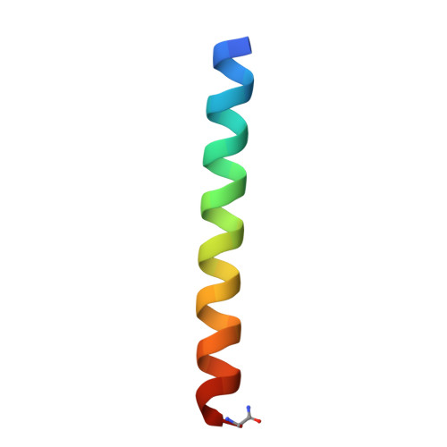
Over the past two decades, designed metallopeptides have held the promise for understanding a variety of fundamental questions in metallobiochemistry; however, these dreams have not yet been realized because of a lack of structural data to elaborate the protein scaffolds before metal complexation and the resultant metalated structures which ultimately exist. This is because there are few reports of structural characterization of such systems either in their metalated or nonmetalated forms and no examples where an apo structure and the corresponding metalated peptide assembly have both been defined by X-ray crystallography. Herein we present X-ray structures of two de novo designed parallel three-stranded coiled coils (designed using the heptad repeat (a → g)) CSL9C (CS = Coil Ser) and CSL19C in their nonmetalated forms, determined to 1.36 and 2.15 A resolutions, respectively. Leucines from either position 9 (a site) or 19 (d site) are replaced by cysteine to generate the constructs CSL9C and CSL19C, respectively, yielding thiol-rich pockets at the hydrophobic interior of these peptides, suitable to bind heavy metals such as As(III), Hg(II), Cd(II), and Pb(II). We use these structures to understand the inherent structural differences between a and d sites to clarify the basis of the observed differential spectroscopic behavior of metal binding in these types of peptides. Cys side chains of (CSL9C)(3) show alternate conformations and are partially preorganized for metal binding, whereas cysteines in (CSL19C)(3) are present as a single conformer. Zn(II) ions, which do not coordinate or influence Cys residues at the designed metal sites but are essential for forming X-ray quality crystals, are bound to His and Glu residues at the crystal packing interfaces of both structures. These "apo" structures are used to clarify the changes in metal site organization between metalated As(CSL9C)(3) and to speculate on the differential basis of Hg(II) binding in a versus d peptides. Thus, for the first time, one can establish general rules for heavy metal binding to Cys-rich sites in designed proteins which may provide insight for understanding how heavy metals bind to metallochaperones or metalloregulatory proteins.
Organizational Affiliation:
Department of Chemistry and Life Sciences Institute, University of Michigan, Ann Arbor, Michigan 48109, USA.